

High throughput single nanoparticle spectroscopy
- PMID: 19472989
- PMCID: PMC2730202
- DOI: 10.1021/nn9003346
High throughput single nanoparticle spectroscopy
Abstract
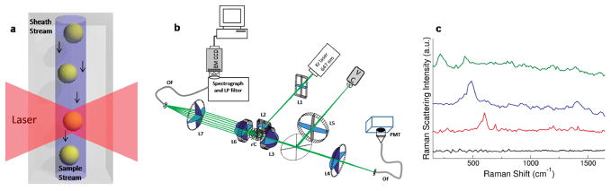
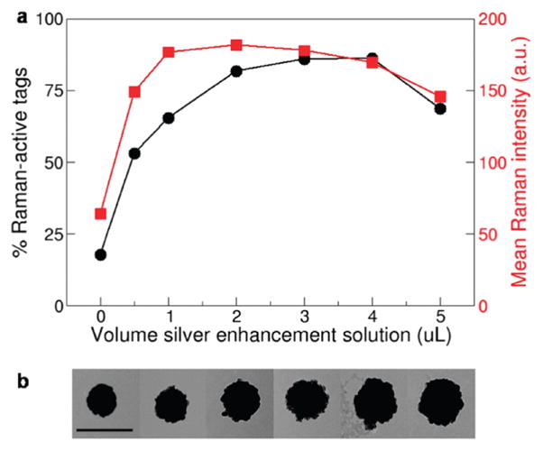
Progress in the development and application of nanoengineered systems is limited by the availability of quantitative measurement techniques. For the engineering of nanoparticle (NP)-based systems, single NP characterization is essential, but existing methods are slow and low throughput. We demonstrate a flow spectroscopy technique capable of analyzing hundreds of nanoparticles per second and use this technique for the high throughput analysis of nanoparticle surface-enhanced resonant Raman scattering (SERRS) tags. By measuring Rayleigh and Raman scattering from thousands of individual tags, tag preparations can be characterized based on their brightness and uniformity. The rapid analysis of individual nanoparticles using high spectral resolution flow spectroscopy will be useful in many areas of nanoengineering.
Figures


Similar articles
-
Single cell analysis using surface enhanced Raman scattering (SERS) tags.Methods. 2012 Jul;57(3):272-9. doi: 10.1016/j.ymeth.2012.03.024. Epub 2012 Apr 4. Methods. 2012. PMID: 22498143 Free PMC article. Review.
-
High-resolution spectral analysis of individual SERS-active nanoparticles in flow.J Am Chem Soc. 2010 May 5;132(17):6081-90. doi: 10.1021/ja909850s. J Am Chem Soc. 2010. PMID: 20143808 Free PMC article.
-
Cancer imaging using surface-enhanced resonance Raman scattering nanoparticles.Nat Protoc. 2017 Jul;12(7):1400-1414. doi: 10.1038/nprot.2017.031. Epub 2017 Jun 22. Nat Protoc. 2017. PMID: 28686581 Free PMC article.
-
Universal surface-enhanced Raman tags: individual nanorods for measurements from the visible to the infrared (514-1064 nm).ACS Nano. 2014 Aug 26;8(8):8600-9. doi: 10.1021/nn503311d. Epub 2014 Aug 14. ACS Nano. 2014. PMID: 25106075
-
Raman Scattering-Based Biosensing: New Prospects and Opportunities.Biosensors (Basel). 2021 Dec 13;11(12):512. doi: 10.3390/bios11120512. Biosensors (Basel). 2021. PMID: 34940269 Free PMC article. Review.
Cited by
-
Raman cell sorting for single-cell research.Front Bioeng Biotechnol. 2024 May 20;12:1389143. doi: 10.3389/fbioe.2024.1389143. eCollection 2024. Front Bioeng Biotechnol. 2024. PMID: 38832129 Free PMC article. Review.
-
Dynamic Imaging Analysis of SERS-Active Nanoparticle Clusters in Suspension.J Phys Chem C Nanomater Interfaces. 2010 Oct 28;114(42):18115-18120. doi: 10.1021/jp107559x. Epub 2010 Oct 5. J Phys Chem C Nanomater Interfaces. 2010. PMID: 23710264 Free PMC article.
-
A trigger channel threshold artifact in nanoparticle analysis.Cytometry A. 2013 Mar;83(3):301-5. doi: 10.1002/cyto.a.22255. Epub 2013 Jan 18. Cytometry A. 2013. PMID: 23335161 Free PMC article.
-
Spectral flow cytometry.Curr Protoc Cytom. 2013 Jan;Chapter 1:1.27.1-1.27.13. doi: 10.1002/0471142956.cy0127s63. Curr Protoc Cytom. 2013. PMID: 23292705 Free PMC article. Review.
-
Single cell analysis using surface enhanced Raman scattering (SERS) tags.Methods. 2012 Jul;57(3):272-9. doi: 10.1016/j.ymeth.2012.03.024. Epub 2012 Apr 4. Methods. 2012. PMID: 22498143 Free PMC article. Review.
References
-
- Cao YWC, Jin RC, Mirkin CA. Nanoparticles with Raman Spectroscopic Fingerprints for DNA and RNA Detection. Science. 2002;297:1536–1540. - PubMed
-
- El-Sayed IH, Huang XH, El-Sayed MA. Surface Plasmon Resonance Scattering and Absorption of Anti-EGFR Antibody Conjugated Gold Nanoparticles in Cancer Diagnostics: Applications in Oral Cancer. Nano Lett. 2005;5:829–834. - PubMed
-
- Haes AJ, Chang L, Klein WL, Van Duyne RP. Detection of a Biomarker for Alzheimer’s Disease from Synthetic and Clinical Samples Using a Nanoscale Optical Biosensor. J Am Chem Soc. 2005;127:2264–2271. - PubMed
-
- Sonnichsen C, Reinhard BM, Liphardt J, Alivisatos AP. A Molecular Ruler Based on Plasmon Coupling of Single Gold and Silver Nanoparticles. Nat Biotechnol. 2005;23:741–745. - PubMed
-
- Loo C, Lowery A, Halas N, West J, Drezek R. Immunotargeted Nanoshells for Integrated Cancer Imaging and Therapy. Nano Lett. 2005;5:709–711. - PubMed
Publication types
MeSH terms
Grants and funding
LinkOut - more resources
Full Text Sources
Other Literature Sources
Miscellaneous
