. 2009;11(3):R32.
doi: 10.1186/bcr2317. Epub 2009 May 27.

WNT signaling enhances breast cancer cell motility and blockade of the WNT pathway by sFRP1 suppresses MDA-MB-231 xenograft growth

Affiliations

WNT signaling enhances breast cancer cell motility and blockade of the WNT pathway by sFRP1 suppresses MDA-MB-231 xenograft growth

Yutaka Matsuda et al. Breast Cancer Res. 2009.

Abstract

Introduction: In breast cancer, deregulation of the WNT signaling pathway occurs by autocrine mechanisms. WNT ligands and Frizzled receptors are coexpressed in primary breast tumors and cancer cell lines. Moreover, many breast tumors show hypermethylation of the secreted Frizzled-related protein 1 (sFRP1) promoter region, causing low expression of this WNT antagonist. We have previously shown that the WNT pathway influences proliferation of breast cancer cell lines via activation of canonical signaling and epidermal growth factor receptor transactivation, and that interference with WNT signaling reduces proliferation. Here we examine the role of WNT signaling in breast tumor cell migration and on xenograft outgrowth.

Methods: The breast cancer cell line MDA-MB-231 was used to study WNT signaling. We examined the effects of activating or blocking the WNT pathway on cell motility by treatment with WNT ligands or by ectopic sFPR1 expression, respectively. The ability of sFRP1-expressing MDA-MB-231 cells to grow as xenografts was also tested. Microarray analyses were carried out to identify targets with roles in MDA-MB-231/sFRP1 tumor growth inhibition.

Results: We show that WNT stimulates the migratory ability of MDA-MB-231 cells. Furthermore, ectopic expression of sFRP1 in MDA-MB-231 cells blocks canonical WNT signaling and decreases their migratory potential. Moreover, the ability of MDA-MB-231/sFRP1-expressing cells to grow as xenografts in mammary glands and to form lung metastases is dramatically impaired. Microarray analyses led to the identification of two genes, CCND1 and CDKN1A, whose expression level is selectively altered in vivo in sFRP1-expressing tumors. The encoded proteins cyclin D1 and p21Cip1 were downregulated and upregulated, respectively, in sFRP1-expressing tumors, suggesting that they are downstream mediators of WNT signaling.

Conclusions: Our results show that the WNT pathway influences multiple biological properties of MDA-MB-231 breast cancer cells. WNT stimulates tumor cell motility; conversely sFRP1-mediated WNT pathway blockade reduces motility. Moreover, ectopic sFRP1 expression in MDA-MB-231 cells has a strong negative impact on tumor outgrowth and blocked lung metastases. These results suggest that interference with WNT signaling using sFRP1 to block the ligand- receptor interaction may be a valid therapeutic approach in breast cancer.


Figures

Figure 1
Figure 1
Ectopic expression of sFRP1 in MDA-MB-231 breast cancer cells. (a) Western analysis was performed on lysates of three MDA-MB-231/secreted Frizzled-related protein 1 (sFRP1) clones and three MDA-MB-231/control clones and the levels of active β-catenin, total β-catenin, α-tubulin and DVL1 to DVL3 determined with specific antisera. Myc-tagged sFRP1 was detected with a Myc-specific antiserum. Lower panel: level of total ERK1/2 and P-ERK1/2 in a MDA-MB-231/sFRP1 clone and a MDA-MB-231/control clone. (b) Activity of the WNT/β-catenin pathway was measured using the TOPFlash T-cell factor reporter system. MDA-MB-231/sFRP1-P1 and control-P1 cells were transiently co-transfected with the TOPFlash reporter plasmid and a pRL-CMV control plasmid, and the reporter activity was measured 48 hours later with a luminometer. y axis, TOPFlash reporter activity normalized by pRL-CMV control activity (arbitrary units), average ± standard error. *P < 0.05. (c) Level of Myc-tagged sFRP1 in two pools of MDA-MB-231/sFRP1 cells determined by western analysis using a Myc-specific antiserum. α-Tubulin levels served as a control. P1 is a mixture of the three clones shown in (a); P2 was generated from > 100 sFRP1-infected clones. (d) MDA-MB-231/sFRP1-P1 and (e) MDA-MB-231/sFRP1-P2 and control-P1 and control-P2 were seeded on six-well dishes (200,000 cells/well) in DMEM, 10% FCS. After 1 day and 3 days, three wells per condition were counted and the average cell numbers were calculated ± standard error of the mean. **P < 0.01, *P < 0.05, n.s. = not significant.
Figure 2
Figure 2
Ectopic expression of sFRP1 in MDA-MB-231 cells suppresses in vivo tumor formation. (a), (b) MDA-MB-231/secreted Frizzled-related protein 1 (sFRP1)-P1 cells and control cells (1 × 10⁶) were injected into mammary fat pads of the indicated number of Balb/c nude mice, and the tumor formation and growth were monitored: (a) average tumor volume, P < 0.01 (two-way repeated-measures analysis of variance (RM ANOVA)); (b) percentage of tumor-free mice on the indicated days after injection, P = 0.0106 (log-rank test). (c) Lysates prepared from individual tumors at the end of the experiment were monitored for Myc-tagged sFRP1 (upper panel) and for p-DVL3 and DVL3 (lower panel) by western analyses. (d) Three independent xenograft experiments using MDA-MB-231/sFRP1-P1 cells (n = 19) and control-P1 cells (n = 20) were performed and data were pooled to calculate the percentage of tumor-free mice 39 days after the injection. (e) Data from the indicated number of mice generated in two independent xenograft experiments with MDA-MB-231/sFRP1-P1 cells and control-P1 cells ((a) and Additional data file 2, Figure 2a) were pooled to yield the tumor growth curve, P < 0.0001 (two-way RM ANOVA). (f), (g) MDA-MB-231/sFRP1-P2 cells and control P2 cells (1 × 10⁶) were injected into mammary fat pads of the indicated number of Balb/c nude mice, and tumor formation and growth were monitored: (f) average tumor volume, *P < 0.01 on day 19 (Student's t test); (g) percentage of tumor-free mice on the indicated days after injection, P = 0.026 (log-rank test). (a), (d), (e) Tumor growth curves shown as the average tumor volume ± standard error.
Figure 3
Figure 3
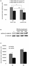
WNT promotes MDA-MB-231 cell migration. (a) Confluent monolayers of MDA-MB-231/secreted Frizzled-related protein 1 (sFRP1)-P1 cells and control-P1 cells were scratched, the medium was changed to Wnt1 conditioned medium (CM) or control CM, and 3 hours later the recovered areas were calculated on nine randomly chosen wound edges. Results presented in arbitrary units. Average recovered areas calculated ± standard error of the mean (SEM). **P < 0.01, n.s. = not significant (P > 0.05). (b) MDA-MB-231 cells were treated with 100 ng/ml recombinant Wnt3a for the indicated time. Active β-catenin was detected by western analysis using specific antiserum; α-tubulin level serves as a loading standard. (c) Confluent monolayers of MDA-MB-231 cells cultured in DMEM, 10% FCS were changed to DMEM, 10% FCS with or without 100 ng/ml recombinant Wnt3a, and 90 minutes later the monolayer surface was scratched. The recovered area was measured on nine randomly chosen wound edges, 3 and 6 hours later. y axis, recovered area (arbitrary unit) ± SEM. **P < 0.01.
Figure 4
Figure 4
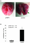
sFRP1 inhibits the metastatic potential of MDA-MB-231 tumor cells. MDA-MB-231/secreted Frizzled-related protein 1 (sFRP1)-P2 cells or control-P2 cells (1 × 10⁶) were injected into five and six Balb/c nude mice, respectively, through the tail vein; 53 days later the mice were sacrificed and the lungs were dissected. (a) Representative picture of lungs from the mice injected with MDA-MB-231/sFRP1-P2 cells (left) and control-P2 cells (right); arrow, metastatic lesion. (b) The total number of lung surface metastases was determined for all the mice and the average is shown ± standard error of the mean. **P < 0.01.
Figure 5
Figure 5
Analysis of proliferation and tumor angiogenesis in MDA-MB-231/sFRP1 xenografts. (a) Mice bearing MDA-MB-231/secreted Frizzled-related protein 1 (sFRP1)-P1 or control-P1 tumors were injected with bromodeoxyuridine (BrdU) and sacrificed 2 hours later. Tumors of similar weight from each group were sectioned and stained with anti-BrdU antibody. Bar = 250 μm. (b) BrdU-positive nuclei and total nuclei were counted in nine randomly chosen areas from each section. Bar graph shows the quantification ± standard error of the mean (SEM). **P < 0.01. (c) Aniokis assays were performed by seeding cells in 1% FCS-containing medium on polyHema-coated plates to prevent adhesion. Cells were harvested 24 hours later, stained with propidium iodide and analyzed with a FACScalibur. Representative cell cycle distribution of three independent control and sFRP1-expressing MDA-MB-231 clones shown. (d) Total blood vessels in tumor sections visualized by staining for the endothelial cell marker CD31. y axis, ratio of CD31+ area/total area. Average calculated from five sFRP1-expressing and eight control tumors ± SEM. n.s., not significant. (e) Functional blood vessels in tumor-bearing mice visualized by injecting FITC-lectin into tail veins 5 minutes before sacrificing. The FITC+ and CD31+ areas were measured on tumor sections and the average was calculated on three sFRP1-expressing and five control tumors. y axis, FITC+ area/CD31+ area ratio ± SEM. n.s., not significant. (d), (e) Image analysis performed using IMARIS software (Bitplane).
Figure 6
Figure 6
Microarray analysis comparing gene expression profiles generated from tumors and from in vitro cultured cells. (a) y axis, 135 probesets (106 genes) showing low expression in secreted Frizzled-related protein 1 (sFRP1)-positive tumors and moderate-high expression in control tumors, in in vitro cultured sFRP1-positive and in control cells. (b) y axis, 84 probesets (62 genes) showing high expression in sFRP1-positive tumors, and low expression in control tumors, in in vitro cultured sFRP1-positive and in control cells. (a), (b) Left to right, columns represent six sFRP1-positive tumors, five control tumors, three MDA-MB-231/sFRP1 clones and three MDA-MB-231/control clones (from Figure 1a). Genes with high, moderate and low expression are indicated in red, black and green, respectively. (c) Normalized gene expression of CCND1 and CDKN1A in MDA-MB-231/sFRP1-P1 and control-P1 tumors, and in MDA-MB-231/sFRP1-P1 and control P1 in vitro cultured cells.
Figure 7
Figure 7
Detection of cyclin D1 and p21Cip1 in tumors and in cultured cells. (a) Tumor sections from mice bearing MDA-MB-231/secreted Frizzled-related protein 1 (sFRP1)-P1 and control tumors were stained with cyclin D1-specific antiserum. Signal intensities, shown in the bar graph, reflect data collected from six MDA-MB-231/sFRP1-P1 tumors and eight control tumors ± standard error of the mean (SEM). *P < 0.05. (b) Cyclin D1 detected in lysates from in vitro cultured MDA-MB-231/sFRP1-P1 cells and control cells by immunoblotting. Bar graph: levels were quantified from three MDA-MB-231/sFRP1 and three control clones (from Figure 1a). Average ± SEM, n.s. = not significant. (c) p21Cip1 detected in tumor lysates from five MDA-MB-231/sFRP1-P1 tumors and three control tumors. (d) Lysates from in vitro cultured MDA-MB-231/sFRP1-P1 and control-P1 cells had no detectable p21Cip1. Lysates from MCF7 control and siRNA-mediated p21Cip1 knockdown tumor cells served as positive and negative controls, respectively; α-tubulin served as loading control.

Comment in

Similar articles

Cited by

References

    1. Clevers H. Wnt/beta-catenin signaling in development and disease. Cell. 2006;127:469–480. doi: 10.1016/j.cell.2006.10.018. - DOI - PubMed
    1. van Amerongen R, Mikels A, Nusse R. Alternative wnt signaling is initiated by distinct receptors. Sci Signal. 2008;1:re9. doi: 10.1126/scisignal.135re9. - DOI - PubMed
    1. Howe LR, Brown AM. Wnt signaling and breast cancer. Cancer Biol Ther. 2004;3:36–41. - PubMed
    1. Katoh M, Katoh M. WNT signaling pathway and stem cell signaling network. Clin Cancer Res. 2007;13:4042–4045. doi: 10.1158/1078-0432.CCR-06-2316. - DOI - PubMed
    1. Huguet EL, McMahon JA, McMahon AP, Bicknell R, Harris AL. Differential expression of human Wnt genes 2, 3, 4, and 7B in human breast cell lines and normal and disease states of human breast tissue. Cancer Res. 1994;54:2615–2621. - PubMed

Publication types