

Aberrant gene expression profile in a mouse model of endometriosis mirrors that observed in women
- PMID: 19473656
- PMCID: PMC2904074
- DOI: 10.1016/j.fertnstert.2009.03.086
Aberrant gene expression profile in a mouse model of endometriosis mirrors that observed in women
Abstract
Objective: To define the altered gene expression profile of endometriotic lesions in a mouse model of surgically induced endometriosis.
Design: Autologous experimental mouse model.
Setting: Medical school department.
Animal(s): Adult C57Bl6 mice.
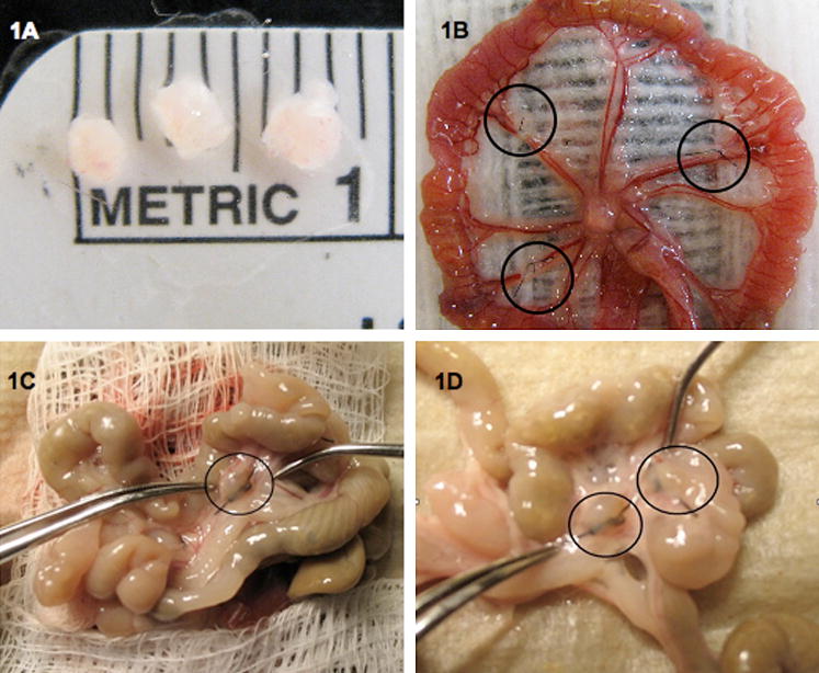
Intervention(s): Endometriosis was surgically induced by autotransplantation of uterine tissue to the intestinal mesentery. Endometriotic lesions and eutopic uteri were recovered at 3 or 29 days after induction.
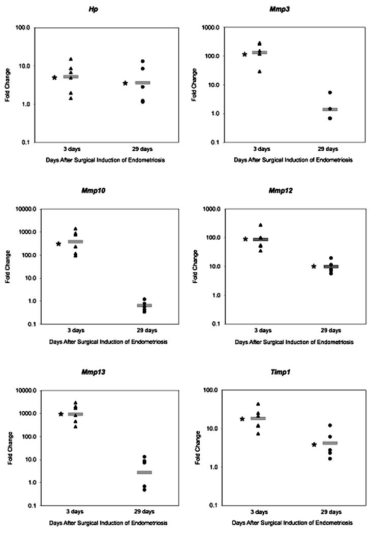
Main outcome measure(s): Altered gene expression was measured in the endometriotic lesion relative to the eutopic uterus by genome-wide complementary DNA microarray analysis and was confirmed by real-time reverse transcriptase-polymerase chain reaction for six genes. Relevant categories of altered genes were identified using gene ontology analysis to determine groups of genes enriched for altered expression.
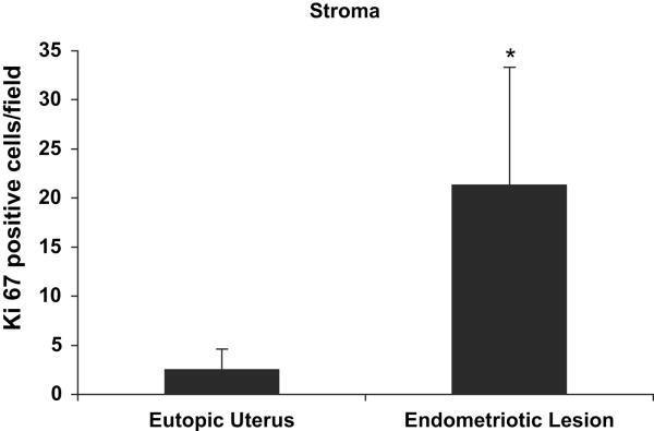
Result(s): The expression of 479 and 114 genes was altered in the endometriotic lesion compared with the eutopic uterus at 3 or 29 days after induction, respectively. Gene ontology enrichment analysis revealed that genes associated with the extracellular matrix, cell adhesions, immune function, cell growth, and angiogenesis were altered in the endometriotic lesion compared with the eutopic uterus.
Conclusion(s): According to gene expression analysis, the mouse model of surgically induced endometriosis is a good model for studying the pathophysiology and treatment of endometriosis.
Copyright 2010 American Society for Reproductive Medicine. Published by Elsevier Inc. All rights reserved.
Conflict of interest statement
Figures
Similar articles
-
Gene expression profiling of the rat endometriosis model.Am J Reprod Immunol. 2007 Oct;58(4):330-43. doi: 10.1111/j.1600-0897.2007.00507.x. Am J Reprod Immunol. 2007. PMID: 17845203
-
miR-451 deficiency is associated with altered endometrial fibrinogen alpha chain expression and reduced endometriotic implant establishment in an experimental mouse model.PLoS One. 2014 Jun 17;9(6):e100336. doi: 10.1371/journal.pone.0100336. eCollection 2014. PLoS One. 2014. PMID: 24937656 Free PMC article.
-
Estrogen receptor beta and matrix metalloproteinase 1 are coexpressed in uterine endometrium and endometriotic lesions of patients with endometriosis.Fertil Steril. 2005 Oct;84 Suppl 2:1249-56. doi: 10.1016/j.fertnstert.2005.06.014. Fertil Steril. 2005. PMID: 16210018
-
Differentially expressed genes in eutopic and ectopic endometrium of women with endometriosis.Fertil Steril. 2010 Apr;93(6):1750-73. doi: 10.1016/j.fertnstert.2008.12.058. Epub 2009 Feb 6. Fertil Steril. 2010. PMID: 19200988
-
Molecular profiling of experimental endometriosis identified gene expression patterns in common with human disease.Fertil Steril. 2007 May;87(5):1180-99. doi: 10.1016/j.fertnstert.2006.07.1550. Fertil Steril. 2007. PMID: 17478174 Free PMC article.
Cited by
-
Exploring the Role of MicroRNAs in Progesterone and Estrogen Receptor Expression in Endometriosis.Biomedicines. 2024 Sep 28;12(10):2218. doi: 10.3390/biomedicines12102218. Biomedicines. 2024. PMID: 39457531 Free PMC article.
-
Distinct spatiotemporal expression of serine proteases Prss23 and Prss35 in periimplantation mouse uterus and dispensable function of Prss35 in fertility.PLoS One. 2013;8(2):e56757. doi: 10.1371/journal.pone.0056757. Epub 2013 Feb 22. PLoS One. 2013. PMID: 23451081 Free PMC article.
-
L1 cell adhesion molecule as a potential therapeutic target in murine models of endometriosis using a monoclonal antibody approach.PLoS One. 2013 Dec 4;8(12):e82512. doi: 10.1371/journal.pone.0082512. eCollection 2013. PLoS One. 2013. PMID: 24324802 Free PMC article.
-
The Pathogenesis of Endometriosis: Molecular and Cell Biology Insights.Int J Mol Sci. 2019 Nov 10;20(22):5615. doi: 10.3390/ijms20225615. Int J Mol Sci. 2019. PMID: 31717614 Free PMC article. Review.
-
A Preliminary Investigation of the Roles of Endometrial Cells in Endometriosis Development via In Vitro and In Vivo Analyses.Int J Mol Sci. 2024 Mar 30;25(7):3873. doi: 10.3390/ijms25073873. Int J Mol Sci. 2024. PMID: 38612685 Free PMC article.
References
-
- Giudice LC, Kao LC. Endometriosis. Lancet. 2004;364:1789–99. - PubMed
-
- Cramer DW, Missmer SA. The epidemiology of endometriosis. Ann N Y Acad Sci. 2002;955:11–22. discussion 34-6, 396-406. - PubMed
-
- Sampson JA. Peritoneal endometriosis due to the menstrual dissemination of endometrial tisse into the peritoneal cavity. Am J Obstet Gynecol. 1927;14 - PubMed
-
- Kruitwagen RF, Poels LG, Willemsen WN, de Ronde IJ, Jap PH, Rolland R. Endometrial epithelial cells in peritoneal fluid during the early follicular phase. Fertil Steril. 1991;55:297–303. - PubMed
-
- Bartosik D, Jacobs SL, Kelly LJ. Endometrial tissue in peritoneal fluid. Fertil Steril. 1986;46:796–800. - PubMed
Publication types
MeSH terms
Grants and funding
LinkOut - more resources
Full Text Sources
Other Literature Sources
Medical
