

Smaller superior temporal gyrus volume specificity in schizotypal personality disorder
- PMID: 19473820
- PMCID: PMC2782902
- DOI: 10.1016/j.schres.2009.04.027
Smaller superior temporal gyrus volume specificity in schizotypal personality disorder
Abstract
Background: Superior temporal gyrus (STG/BA22) volume is reduced in schizophrenia and to a milder degree in schizotypal personality disorder (SPD), representing a less severe disorder in the schizophrenia spectrum. SPD and Borderline personality disorder (BPD) are severe personality disorders characterized by social and cognitive dysfunction. However, while SPD is characterized by social withdrawal/anhedonia, BPD is marked by hyper-reactivity to interpersonal stimuli and hyper-emotionality. This is the first morphometric study to directly compare SPD and BPD patients in temporal lobe volume.
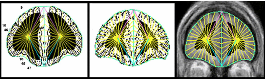
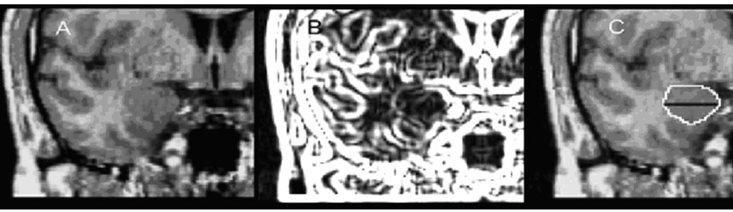
Methods: We compared three age-, sex-, and education-matched groups: 27 unmedicated SPD individuals with no BPD traits, 52 unmedicated BPD individuals with no SPD traits, and 45 healthy controls. We examined gray matter volume of frontal and temporal lobe Brodmann areas (BAs), and dorsal/ventral amygdala from 3-T magnetic resonance imaging.
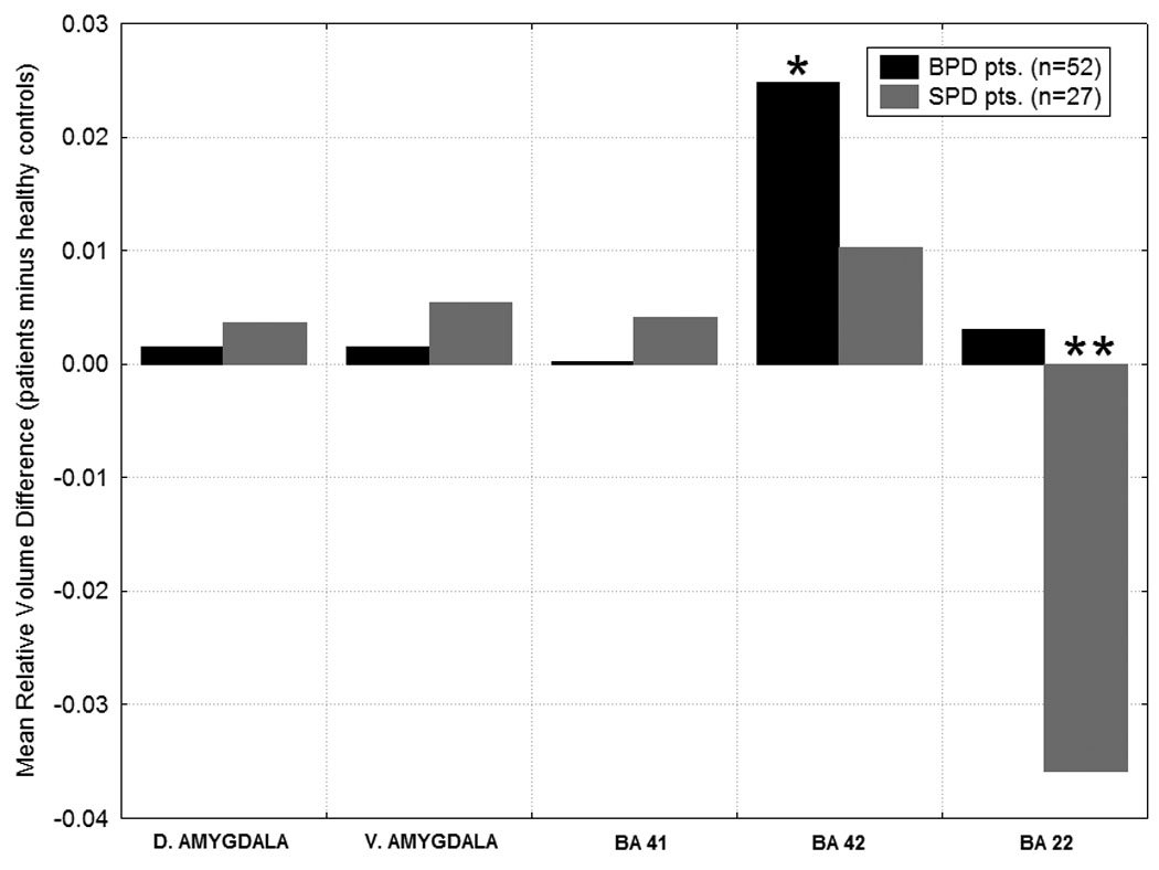
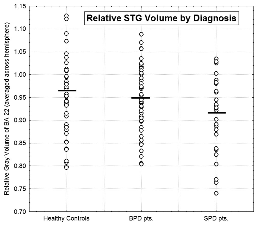
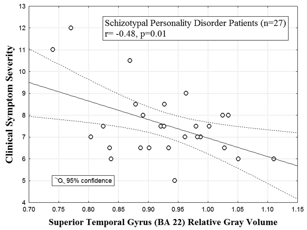
Results: In the STG, an auditory association area reported to be dysfunctional in SPD and BPD, the SPD patients had significantly smaller volume than healthy controls and BPD patients. No group differences were found between BPD patients and controls. Smaller BA22 volume was associated with greater symptom severity in SPD patients. Reduced STG volume may be an important endophenotype for schizophrenia-spectrum disorders. SPD is distinct from BPD in terms of STG volume abnormalities which may reflect different underlying pathophysiological mechanisms and could help discriminate between them.
Conflict of interest statement
There are no conflicts of interest for any of the authors.
Figures
Similar articles
-
Reduced anterior and posterior cingulate gray matter in borderline personality disorder.Biol Psychiatry. 2005 Oct 15;58(8):614-23. doi: 10.1016/j.biopsych.2005.04.029. Epub 2005 Jul 5. Biol Psychiatry. 2005. PMID: 15993861
-
Schizotypal personality disorder and MRI abnormalities of temporal lobe gray matter.Biol Psychiatry. 1999 Jun 1;45(11):1393-402. doi: 10.1016/s0006-3223(99)00030-x. Biol Psychiatry. 1999. PMID: 10356620 Free PMC article.
-
Cingulate and temporal lobe fractional anisotropy in schizotypal personality disorder.Neuroimage. 2011 Apr 1;55(3):900-8. doi: 10.1016/j.neuroimage.2010.12.082. Epub 2011 Jan 9. Neuroimage. 2011. PMID: 21223999 Free PMC article.
-
A review of structural MRI and diffusion tensor imaging in schizotypal personality disorder.Curr Psychiatry Rep. 2012 Feb;14(1):70-8. doi: 10.1007/s11920-011-0241-z. Curr Psychiatry Rep. 2012. PMID: 22006127 Free PMC article. Review.
-
[Biological markers in schizotypal and borderline personality disorders].Encephale. 2000 Nov-Dec;26(6):42-54. Encephale. 2000. PMID: 11217538 Review. French.
Cited by
-
Children's Brain Development Benefits from Longer Gestation.Front Psychol. 2011 Feb 9;2:1. doi: 10.3389/fpsyg.2011.00001. eCollection 2011. Front Psychol. 2011. PMID: 21713130 Free PMC article.
-
Gamma Oscillations as a Biomarker of Neural Circuit Function in Psychosis: Where Are We, and Where Do We Go from Here?Adv Neurobiol. 2024;40:321-349. doi: 10.1007/978-3-031-69491-2_12. Adv Neurobiol. 2024. PMID: 39562450 Review.
-
Multimodal morphometry and functional magnetic resonance imaging in schizophrenia and auditory hallucinations.World J Radiol. 2012 Apr 28;4(4):159-66. doi: 10.4329/wjr.v4.i4.159. World J Radiol. 2012. PMID: 22590670 Free PMC article.
-
Frontal and temporal cortical volume, white matter tract integrity, and hemispheric asymmetry in schizotypal personality disorder.Schizophr Res. 2018 Jul;197:226-232. doi: 10.1016/j.schres.2018.01.025. Epub 2018 Feb 14. Schizophr Res. 2018. PMID: 29454512 Free PMC article.
-
Auditory steady state response in the schizophrenia, first-degree relatives, and schizotypal personality disorder.Schizophr Res. 2012 Apr;136(1-3):143-9. doi: 10.1016/j.schres.2012.01.003. Epub 2012 Jan 28. Schizophr Res. 2012. PMID: 22285558 Free PMC article.
References
-
- Barbas H. Specialized Elements of Orbitofrontal Cortex in Primates. Vol. 1121. Annals of the New York Academy of Sciences; 2007. pp. 10–32. - PubMed
-
- Binder JR, Rao SM, Hammeke TA, Yetkin FZ, Jesmanowicz A, Bandettini PA, Wong EC, Etskowski LD, Goldstein MD, Haughton VM, Hyde JS. Functional magnetic resonance imaging of human auditory cortex. Annals of Neurology. 1994;35:662–672. - PubMed
-
- Binder J, Frost J, Hammeke T, Bellgowan PS, Springer JA, Kaufman JN, Possing ET. Human temporal lobe activation by speech and nonspeech sounds. Cerebral Cortex. 2000;10:512–528. - PubMed
-
- Brambilla P, Soloff PH, Sala M, Nicoletti MA, Keshavan MS, Soares JC. Anatomical MRI study of borderline personality disorder patients. Psychiatry Research. 2004;131:125–133. - PubMed
-
- Buchsbaum MS, Nenadic I, Hazlett EA, Spiegel-Cohen J, Fleischman MB, Akhavan A, Silverman JM, Siever LJ. Differential metabolic rates in prefrontal and temporal Brodmann areas in schizophrenia and schizotypal personality disorder. Schizophrenia Research. 2002;54:141–150. - PubMed
Publication types
MeSH terms
Grants and funding
LinkOut - more resources
Full Text Sources
