. 2009 Jul 17;284(29):19427-36.
doi: 10.1074/jbc.M109.003442. Epub 2009 May 27.

Cdc42 and the phosphatidylinositol 3-kinase-Akt pathway are essential for PspC-mediated internalization of pneumococci by respiratory epithelial cells

Affiliations

Cdc42 and the phosphatidylinositol 3-kinase-Akt pathway are essential for PspC-mediated internalization of pneumococci by respiratory epithelial cells

Vaibhav Agarwal et al. J Biol Chem. .

Abstract

The pneumococcal surface protein C (PspC) is a major adhesin of Streptococcus pneumoniae, the cause of lobar pneumonia and invasive diseases. PspC interacts in a human-specific manner with the ectodomain of the human polymeric immunoglobulin receptor (pIgR) produced by respiratory epithelial cells. By adopting the retrograde machinery of human pIgR, this protein-protein interaction promotes colonization and transcytosis across the epithelial layer. Here, we explored the role of Rho family guanosine triphosphatases (GTPases), phosphatidylinositol 3-kinase (PI3K) and protein kinase B (Akt) for ingestion of pneumococci via the human pIgR. Inhibition experiments suggested that the host-cell actin microfilaments and microtubules are essential for this pneumococcal uptake mechanism. By using specific GTPase-modifying toxins, inhibitors, and GTPase expression constructs we demonstrate that Cdc42, but not Rac1 and RhoA are involved in PspC-mediated invasion of pneumococci into host cells. Accordingly, Cdc42 is time-dependently activated during ingestion of pneumococci. In addition, PI3K and Akt are essential for ingestion of pneumococci by respiratory epithelial cells via the PspC-pIgR interaction. The subunit p85alpha of PI3K and Akt was activated during the infection process. Moreover, Akt activation upon pneumococcal invasion depends on PI3K. In conclusion, our results illustrate for the first time key signaling molecules of host cells that are required for PspC-pIgR-mediated invasion of pneumococci into epithelial cells. This unique and specific bacterial entry process is dependent on the cooperation and activation of Rho family GTPase Cdc42, PI3K, and Akt.


Figures

FIGURE 1.
FIGURE 1.
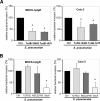
PspC-hpIgR-mediated adherence and invasion of MDCK-hpIgR and Calu-3 cells by wild-type pneumococci and its isogenic pspC-mutant. A, adherence of pneumococci was determined by counting the cfu (colony forming unit) per well obtained from sample aliquots plated onto blood agar plates after 1 h of infection. *, p < 0.02 relative to infections carried out with the wild-type strain. B and C, invasion and intracellular survival of pneumococci were determined by the antibiotic protection assay. *, p < 0.02 relative to infections carried out with the wild-type strain or relative to MDCK-hpIgR. D, the invasion and intracellular survival of pneumococci in host cells were determined in the presence of antibodies recognizing the secretory component of hpIgR (α-SC, 8 μg/well), pre-immune serum, or absence of antibody using the antibiotic protection assay. Invasion of S. pneumoniae in the absence of α-SC was set to 100%. *, p < 0.001 relative to infections carried out in absence of antibodies.
FIGURE 2.
FIGURE 2.
Invasion of pneumococci via pIgR requires the dynamics of the host cell actin cytoskeleton and microtubuli. A, pneumococcal adherence to MDCK-hpIgR and Calu-3 cells was determined in the absence (control, Ctrl) or presence of various inhibitors. Adherence of S. pneumoniae in the absence of an inhibitor was set to 100%. B, invasion of MDCK-hpIgR or Calu-3 cells with pneumococci was followed in the absence (control) or presence of inhibitors of actin filaments and microtubules, including cytochalasin D (CytoD, 125 nm), latrunculin B (LatB, 50 nm), jasplakinolide (Jasp, 100 nm), and nocodazole (Noco, 10 μm), by the antibiotic protection assay. Invasion of S. pneumoniae in the absence of an inhibitor was set to 100%. *, p < 0.001 relative to infections carried out in absence of an inhibitor. C, immunofluorescence microscopy illustrating changes of the actin cytoskeleton after infecting host cells for 3 h with pneumococci. F-actin was stained with AlexaFluor-488 phalloidin, and intracellular pneumococci were stained with AlexaFluor-568 (red), whereas adherent bacteria were stained with Cy5 and hence, bacteria appear pink (blue/red stain). Uninfected host cells (a and d) and infected host cells (b and e), respectively. Higher magnifications (c and e) illustrate changes of the actin cytoskeleton during pIgR mediated infection of MDCK-hpIgR or Calu-3 cells.
FIGURE 3.
FIGURE 3.
Effect of bacterial protein toxins and pharmacological inhibitors targeting small Rho GTPases on PspC-hpIgR-mediated host cell internalization of pneumococci. Pneumococcal invasion in MDCK-hpIgR and Calu-3 cells was determined in the absence (Ctrl) or presence of (A) C. difficile toxin B, TcdB-10463 (30 ng ml−1), TcdB-1470 (100 ng ml−1), and (B) specific individual inhibitors of Rho family GTPases such as Y27632 (50 μm), Rac1 inhibitor NSC23766 (50 μm), or Cdc42 inhibitor secramine A (10 μm) by the antibiotic protection assay. Invasion of S. pneumoniae in the absence of toxin or inhibitor was set to 100%. *, p < 0.05 relative to infections carried out in absence of toxins or inhibitor.
FIGURE 4.
FIGURE 4.
Activity of small GTPases Cdc42 is essential for PspC-hpIgR-mediated invasion of MDCK-hpIgR cells by pneumococci. The effect of expression of dominant-negative (dn) forms of Rho GTPases Rho-T19N, Rac1-T17N, Cdc42-T17N, or constitutively active (ca) Cdc42-Q61L on pneumococcal invasion was assessed by the antibiotic protection assay after the constructs were transiently transfected into MDCK-hpIgR cells. Invasion by S. pneumoniae into vector-transfected host cells (Ctrl) was set to 100%. *, p < 0.001 for dominant-negative Cdc42, and p < 0.03 for constitutively active Cdc42 relative to infections performed with vector-transfected cells.
FIGURE 5.
FIGURE 5.
Time-dependent activation of Cdc42 GTPase during pIgR-mediated pneumococcal invasion. Host cell lysates of MDCK-hpIgR and Calu-3 cells, respectively, were prepared after infection with pneumococci for indicated time points and were employed in pulldown assays of small GTPases (upper panels). GST-PAK was used for Rac1 and Cdc42, whereas GST-mDia was employed for RhoA. Precipitates were separated by 14% SDS-PAGE and analyzed using GTPase-specific antibodies. The pulldown assays from lysates that were prepared from uninfected host cells were used as controls (0 min). Total amounts of GTPases Rac1, Cdc42, or RhoA were analyzed using sample aliquots of lysates (lower panels). Quantification of GTPase activity of Cdc42: 100% of activity corresponds to the highest amount of detected GTPase-GTP levels.
FIGURE 6.
FIGURE 6.
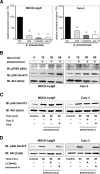
PI3K and Akt are activated during pneumococcal invasion via pIgR. A, invasion and intracellular survival of the bacteria in MDCK-hpIgR and Calu-3 cells were monitored in the absence (Ctrl) or presence of PI3K inhibitors wortmannin (50 nm) or LY294002 (50 μm) and in the presence of Akt Inhibitor VIII (Akti, 10 μm) by the antibiotic protection assay. Pneumococcal invasion in the absence of the inhibitor was set to 100%. *, p < 0.001 relative to infections carried out in the absence of inhibitor. B, pneumococcal infection of MDCK-hpIgR and Calu-3 cells induces phosphorylation of PI3K subunit p85α and Akt. Host cell lysates of MDCK-hpIgR and Calu-3 cells prepared after pneumococcal infections and separated by 10% SDS-PAGE. Activation of kinases were analyzed using antibodies against phosphorylated forms of PI3K subunit p85α (upper panel) or Akt (pAkt) (middle panel). The membrane was stripped and reprobed with total Akt antibody and used as loading control (lower panel). C, activation of Akt is independent of pneumococcal invasion. Pneumococcal invasion was blocked by inhibition of the actin cytoskeleton dynamics with Cytochalasin D (CytoD) or jasplakinolide (Jasp) and activation of Akt followed. Total Akt served as loading control (lower panel). D, Akt is activated in a PI3K-dependent but Cdc42-independent manner. Activation of Akt was followed in the presence of PI3K inhibitor LY294002 (50 μm) or Cdc42 inhibitor secramine A (10 μm). Total Akt served as loading control (lower panel).
FIGURE 7.
FIGURE 7.
Akt and Cdc42 are essential for PspC-mediated pneumococcal uptake by primary respiratory host cells. A, adherence of pneumococci was determined by counting the cfu per well obtained from sample aliquots plated onto blood agar plates after 1 h of host cell infection. *, p < 0.02 relative to infections carried out with the wild-type strain. B, immunofluorescence microscopy of S. pneumoniae wild type (WT) and its isogenic PspC mutant attached to primary human respiratory epithelial cells. C, invasion and intracellular survival of pneumococci were determined by the antibiotic protection assay. *, p < 0.02 relative to infections carried out with the wild-type strain. D, invasion and intracellular survival of the bacteria in primary cells human bronchial epithelial cell cells were determined in the absence (Ctrl) or presence of Akt Inhibitor VIII (Akti 1/2, 10 μm) and the presence of Cdc42 inhibitor secramine A (10 μm) by the antibiotic protection assay. *, p < 0.02 relative to infections carried out in the absence of inhibitor.
FIGURE 8.
FIGURE 8.
Schematic model of pneumococci-induced signaling molecules. Solid arrows depict activated signaling molecules due to invasion of respiratory epithelial cells by pneumococci via the PspC-pIgR interaction. The dotted arrows point to the known implication of these signaling molecules toward modulation of host cell cytoskeleton.

Similar articles

Cited by

References

    1. Cartwright K. (2002) Eur. J. Pediatr. 161, 188–195 - PubMed
    1. Hammerschmidt S., Agarwal V., Kunert A., Haelbich S., Skerka C., Zipfel P. F. (2007) J. Immunol. 178, 5848–5858 - PubMed
    1. Rennemeier C., Hammerschmidt S., Niemann S., Inamura S., Zähringer U., Kehrel B. E. (2007) FASEB J. 21, 3118–3132 - PubMed
    1. Bergmann S., Lang A., Rohde M., Agarwal V., Rennemeier C., Grashoff C., Preissner K. T., Hammerschmidt S. (2009) J. Cell Sci. 122, 256–267 - PubMed
    1. Hammerschmidt S. (2006) Curr. Opin. Microbiol. 9, 12–20 - PubMed

Publication types

MeSH terms

Substances