. 2009 Jul 17;284(29):19340-8.
doi: 10.1074/jbc.M109.004473. Epub 2009 May 27.

Identification of the C1q-binding Sites of Human C1r and C1s: a refined three-dimensional model of the C1 complex of complement

Affiliations

Identification of the C1q-binding Sites of Human C1r and C1s: a refined three-dimensional model of the C1 complex of complement

Isabelle Bally et al. J Biol Chem. .

Abstract

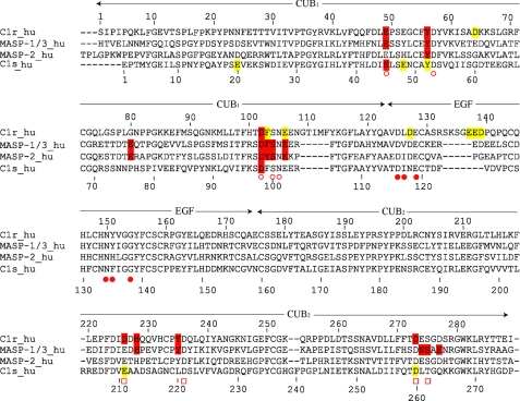
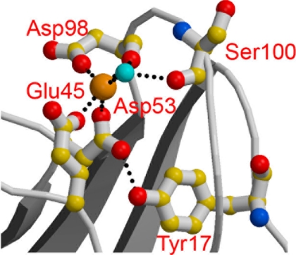
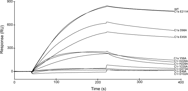
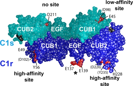
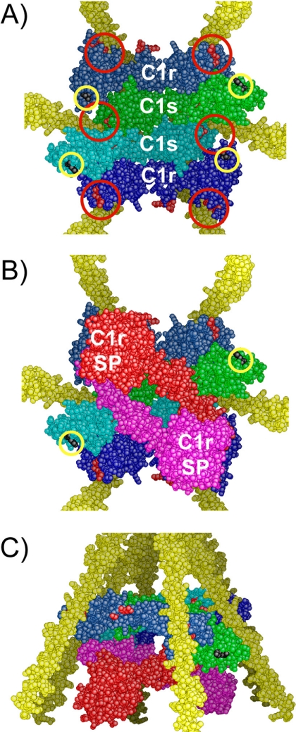
The C1 complex of complement is assembled from a recognition protein C1q and C1s-C1r-C1r-C1s, a Ca(2+)-dependent tetramer of two modular proteases C1r and C1s. Resolution of the x-ray structure of the N-terminal CUB(1)-epidermal growth factor (EGF) C1s segment has led to a model of the C1q/C1s-C1r-C1r-C1s interaction where the C1q collagen stem binds at the C1r/C1s interface through ionic bonds involving acidic residues contributed by the C1r EGF module (Gregory, L. A., Thielens, N. M., Arlaud, G. J., Fontecilla-Camps, J. C., and Gaboriaud, C. (2003) J. Biol. Chem. 278, 32157-32164). To identify the C1q-binding sites of C1s-C1r-C1r-C1s, a series of C1r and C1s mutants was expressed, and the C1q binding ability of the resulting tetramer variants was assessed by surface plasmon resonance. Mutations targeting the Glu(137)-Glu-Asp(139) stretch in the C1r EGF module had no effect on C1 assembly, ruling out our previous interaction model. Additional mutations targeting residues expected to participate in the Ca(2+)-binding sites of the C1r and C1s CUB modules provided evidence for high affinity C1q-binding sites contributed by the C1r CUB(1) and CUB(2) modules and lower affinity sites contributed by C1s CUB(1). All of the sites implicate acidic residues also contributing Ca(2+) ligands. C1s-C1r-C1r-C1s thus contributes six C1q-binding sites, one per C1q stem. Based on the location of these sites and available structural information, we propose a refined model of C1 assembly where the CUB(1)-EGF-CUB(2) interaction domains of C1r and C1s are entirely clustered inside C1q and interact through six binding sites with reactive lysines of the C1q stems. This mechanism is similar to that demonstrated for mannan-binding lectin (MBL)-MBL-associated serine protease and ficolin-MBL-associated serine protease complexes.


Figures

FIGURE 1.
FIGURE 1.
Purification of the recombinant C1s-C1r-C1r-C1s tetramer. The tetramer was reconstituted from wild type C1s and the S637A C1r mutant. SDS-PAGE analysis was performed after ion exchange chromatography (lanes 1 and 2) and after high pressure gel permeation (lanes 3 and 4), under reducing (lanes 1 and 3), and nonreducing conditions (lanes 2 and 4). The arrows indicate the position of recombinant C1r and C1s under both conditions. The positions of reduced and unreduced standard proteins are indicated on the left and right sides of the figure, respectively.
FIGURE 2.
FIGURE 2.
Sequence alignment of selected CUB1-EGF-CUB2 segments. The amino acid numberings of both C1r (top) and C1s (bottom) are shown, and the approximate domain boundaries are indicated. Residues known to contribute Ca2+ ligands in C1s, MAp19, and MASP-1/3 are marked with open circles (CUB1 site), closed circles (EGF site), and open squares (CUB2 site). C1r and C1s residues interacting with C1q, and MASP residues interacting with MBL and ficolins are colored red. Mutations in C1r and C1s having little or no effect on C1 assembly are colored yellow. The implication of C1s Tyr52 in C1q binding cannot be excluded.
FIGURE 3.
FIGURE 3.
Structure of the Ca2+-binding site of the C1s CUB1 module. Oxygen atoms are shown in red, and nitrogen atoms are in blue. The light blue sphere represents a water molecule. Ionic bonds and hydrogen bonds are represented by dotted lines (adapted from Gregory et al. (16)).
FIGURE 4.
FIGURE 4.
Analysis by surface plasmon resonance spectroscopy of the interaction between selected C1s-C1r-C1r-C1s variants and immobilized C1q. C1q was immobilized on the sensor chip as described under “Experimental Procedures.” The native C1s-C1r-C1r-C1s tetramer reconstituted from wild type C1s and the S637A C1r mutant, and selected variants with mutations in either C1r or C1s were injected at a concentration of 10 nm.
FIGURE 5.
FIGURE 5.
Three-dimensional space-filling representation of the C1r/C1s CUB1-EGF-CUB2 heterodimer. The head-to-tail C1r/C1s CUB1-EGF-CUB2 heterodimeric structure was built as described under “Experimental Procedures.” C1r and C1s domains are colored dark blue and turquoise, respectively. The Ca2+-binding sites of the C1r and C1s CUB modules are marked by residues Glu49, Tyr56, and Asp102 (C1r CUB1); Asp226, His228, and Tyr235 (C1r CUB2); Glu45 and Asp98 (C1s CUB1); and Glu211 (C1s CUB2), shown in red. Residues barely visible are indicated in parentheses. The acidic cluster Glu137-Glu-Asp139 in the C1r EGF module (indicated by an asterisk) is also shown in red. The C-terminal ends of the C1r and C1s CUB2 modules are shown in black.
FIGURE 6.
FIGURE 6.
A refined three-dimensional model of C1 assembly. A, bottom view of the assembly between the C1r/C1s CUB1-EGF-CUB2 interaction domains and the C1q collagen stems. C1r and C1s domains are colored dark blue and light blue, respectively. The approximate positioning of the six C1q-binding sites contributed by C1r and C1s is indicated by red circles. The C-terminal ends of the C1r and C1s CUB2 modules are marked by yellow circles. B, bottom view of the assembly featuring the CCP1-CCP2-SP catalytic domains of C1r, shown in red and pink. The C1s catalytic domains, emerging from the C-terminal end of both C1s CUB2 modules (yellow circles), are not shown for clarity. SP, serine protease domain. C, side view of the assembly shown in B.

References

    1. Cooper N. R. (1985) Adv. Immunol. 37, 151–216 - PubMed
    1. Arlaud G. J., Colomb M. G., Gagnon J. (1987) Immunol. Today 8, 106–111 - PubMed
    1. Arlaud G. J., Gaboriaud C., Thielens N. M., Rossi V., Bersch B., Hernandez J. F., Fontecilla-Camps J. C. (2001) Immunol. Rev. 180, 136–145 - PubMed
    1. Kishore U., Reid K. B. M. (2000) Immunopharmacology 49, 159–170 - PubMed
    1. Bork P., Beckmann G. (1993) J. Mol. Biol. 231, 539–545 - PubMed

Publication types

MeSH terms