. 2009 Jul 17;284(29):19392-401.
doi: 10.1074/jbc.M109.023283. Epub 2009 May 27.

Docking of PRAK/MK5 to the atypical MAPKs ERK3 and ERK4 defines a novel MAPK interaction motif

Affiliations

Docking of PRAK/MK5 to the atypical MAPKs ERK3 and ERK4 defines a novel MAPK interaction motif

Espen Aberg et al. J Biol Chem. .

Abstract

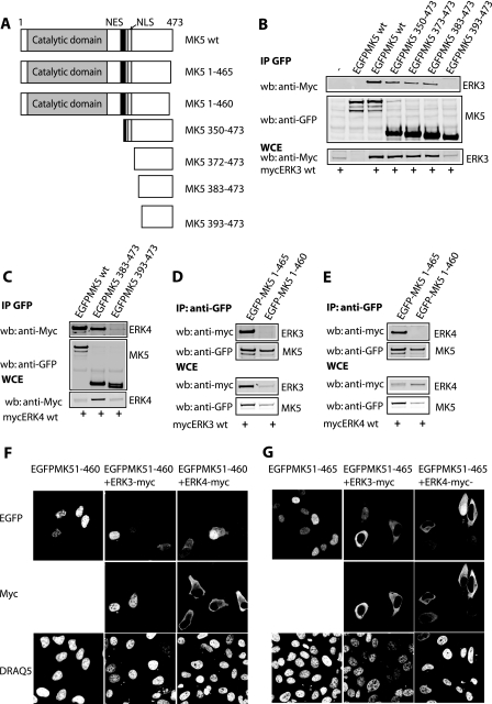
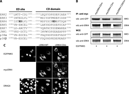
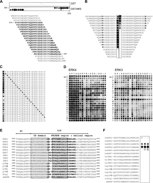
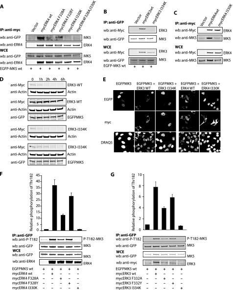
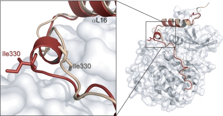
ERK3 and ERK4 are atypical MAPKs in which the canonical TXY motif within the activation loop of the classical MAPKs is replaced by SEG. Both ERK3 and ERK4 bind, translocate, and activate the MAPK-activated protein kinase (MK) 5. The classical MAPKs ERK1/2 and p38 interact with downstream MKs (RSK1-3 and MK2-3, respectively) through conserved clusters of acidic amino acids, which constitute the common docking (CD) domain. In contrast to the classical MAPKs, the interaction between ERK3/4 and MK5 is strictly dependent on phosphorylation of the SEG motif of these kinases. Here we report that the conserved CD domain is dispensable for the interaction of ERK3 and ERK4 with MK5. Using peptide overlay assays, we have defined a novel MK5 interaction motif (FRIEDE) within both ERK4 and ERK3 that is essential for binding to the C-terminal region of MK5. This motif is located within the L16 extension lying C-terminal to the CD domain in ERK3 and ERK4 and a single isoleucine to lysine substitution in FRIEDE totally abrogates binding, activation, and translocation of MK5 by both ERK3 and ERK4. These findings are the first to demonstrate binding of a physiological substrate via this region of the L16 loop in a MAPK. Furthermore, the link between activation loop phosphorylation and accessibility of the FRIEDE interaction motif suggests a switch mechanism for these atypical MAPKs in which the phosphorylation status of the activation loop regulates the ability of both ERK3 and ERK4 to bind to a downstream effector.


Figures

FIGURE 1.
FIGURE 1.
Multiple determinants within the C terminus of MK5 are important for interaction with ERK4. A, schematic representation of the MK5 constructs used is shown. B and C, EGFP co-immunoprecipitation (IP) assays were performed on lysates from HeLa cells co-transfected with the indicated expression vectors. Bound ERK3 (B) or ERK4 (C) was detected using an anti-Myc antibody (top panel). EGFPMK5 was detected with an anti-GFP antibody (middle panel). Equal expression of ERK3 and ERK4 was verified by Western blotting (wb) of the whole cell extract (WCE) with anti-Myc antibody (bottom panel). D and E, HeLa cells were co-transfected with vectors encoding either Myc-ERK3 (D) or Myc-ERK4 (E) and EGFP-MK5 1–465 or EGFP-MK5 1–460. After 24 h, whole cell extracts were prepared and ERK4 was immunoprecipitated with an anti-GFP antibody. Co-immunoprecipitated ERK3 (D) or ERK4 (E) was detected by Western blot analysis using an anti-Myc antibody (top panel), whereas MK5 was detected using a polyclonal anti-GFP antibody (second panel). Equal expression of ERK3 and ERK4 or EGFP-MK5 were verified by Western blotting of the whole cell extract with anti-Myc antibody (third panel) or anti-GFP antibody (bottom panel). F and G, HeLa cells were co-transfected with expression vectors encoding EGFP-MK5 1–460 or EGFP-MK5 1–465 and mycERK3 (F) or Myc-ERK4 (G). After 24 h, the cells were fixed, and EGFP-MK5 was visualized directly (top panel), and ERK3 or ERK4 was visualized by staining with an anti-Myc antibody and an Alexa 594-coupled secondary (anti-mouse) antibody (middle panel). The cell nuclei were visualized by DRAQ5 staining (bottom panel).
FIGURE 2.
FIGURE 2.
The CD domain of ERK4 is dispensable for its ability to interact with MK5. A, amino acid sequences in the CD domains for different human members of the MAPK family are shown. Residues in bold type indicate surface-exposed and negatively charged amino acids within the CD domain that are close to one another in the tertiary structure (20). Bold type indicate mutations introduced by us within the ERK4 CD domain (ERK4 CDm) and ED site (ERK4 CDEDm). B, HeLa cells were co-transfected with EGFP-MK5 and Myc-ERK4 WT, Myc-ERK4 CDm, or Myc-ERK4 CDEDm. After 24 h, the cells were lysed, and Myc-ERK4 was immunoprecipitated (IP) using an anti-Myc antibody. The immunoprecipitates were then analyzed by SDS-PAGE and Western blotting (wb) using either an anti-GFP antibody against MK5 (top panel) or an anti-ERK4 antibody (second panel). Equal expression of EGFP-MK5 and ERK4 were verified by Western blotting of the whole cell extract (WCE) with either an anti-GFP antibody (third panel) or anti-Myc antibody (bottom panel). C, HeLa cells were co-transfected with Myc-ERK4 WT or Myc-ERK4 CDm. After 24 h, the cells were fixed, and EGFP-MK5 was visualized directly (top panel), and ERK4 was visualized by staining with an anti-Myc antibody and an Alexa 594-coupled secondary (anti-mouse) antibody (middle panel). The cell nuclei were visualized by DRAQ5 staining (bottom panel).
FIGURE 3.
FIGURE 3.
Identification of the FRIEDE motif within the L16 loop of ERK4 as an MK5 binding determinant. In all overlay assays, the indicated peptide sequences were bound on cellulose membranes and probed with 1 μg/ml GST or GST-MK5 for 2 h and detected with an anti-GST antibody conjugated to horseradish peroxidase. Overlay assay with GST alone gave little or no signal in all experiments (A and data not shown). A, a sequence corresponding to amino acids 292–370 was spotted as 20-mer peptides on a cellulose filter as a 1-amino acid shifted array covering the entire sequence. Positive spots correspond to the highlighted sequences. B, increasing N- and C-terminal truncations of the ERK4 sequence were spotted and evaluated for MK-5 binding. Amino acids defining the binding boundaries are highlighted. C, the ERK4 sequence was subjected to an alanine scan by sequential substitution to identify critical amino acids for binding to MK-5. D, a two-dimensional peptide array was constructed by replacing the 18-residue sequence in ERK4 (left panel) and the homologous region of ERK3 (right panel) with all 20 amino acids as indicated. E, alignment of the kinase domain XI and L16 including the CD domain and the FRIEDE motif for the indicated MAPKs is shown. F, to test the specificity of MK5 for the FRIEDE motif in ERK3 and ERK4, we spotted the corresponding sequences of the indicated MAPKs in triplicates on a filter and probed with GST-MK5.
FIGURE 4.
FIGURE 4.
Mutations in the FRIEDE motif affect the ability of both ERK3 and ERK4 to interact with, translocate, and activate MK5. A, HeLa cells were transfected with the indicated versions of Myc-tagged ERK4 and WT EGFP-MK5. After 24 h, the cells were lysed, and Myc-ERK4 was immunoprecipitated (IP) using an anti-Myc antibody. The immunoprecipitates were analyzed by SDS-PAGE and Western blotting (wb) using an anti-GFP antibody for MK5 (top panel), an anti-ERK4 antibody for ERK4 (second panel). B, HeLa cells were co-transfected with vectors encoding Myc-ERK3 WT or Myc-ERK3 I334K and EGFP-MK5 WT. After 24 h, whole cell extracts were prepared, and EGFP-MK5 was immunoprecipitated with an anti-GFP antibody. ERK3 and EGFP-MK5 were detected by Western blotting using an anti-Myc antibody or a polyclonal anti-GFP antibody, respectively. C, HeLa cells were transfected with the indicated vectors. After 24 h, whole cell extracts were prepared, and Myc-tagged ERK4 was immunoprecipitated with an EZview anti c-Myc affinity gel. The immunoprecipitates were then analyzed by SDS-PAGE and Western blotting using an anti-Myc antibody to detect ERK4 and a monoclonal antibody against MK5. D, HeLa cells were transfected with Myc-ERK3-WT or Myc-ERK3 I334K alone or together with EGFP-MK5 WT. After 24 h, cycloheximide (10 μg/ml) was added, and the cells were harvested at the indicated time. ERK3 were visualized by Western blotting using an anti-Myc antibody. The blots were also probed with anti-actin antibody to ensure equal loading and anti-GFP antibody to verify expression of EGFP-MK5. E, HeLa cells were transfected with expression vectors encoding either EGFP-MK5 alone or in combination with either Myc-ERK3 WT, Myc-ERK3 I334K Myc-ERK4 WT, and Myc-ERK4 I330K. After 24 h the cells were fixed, and EGFP-MK5 was visualized directly (top panel). ERK3 and ERK4 were visualized by staining with an anti-Myc antibody and Alexa 594 anti-mouse antibody (middle panel). The nuclei were visualized by DRAQ5 staining (bottom panel). F and G, HeLa cells were co-transfected with the indicated expression plasmids encoding the indicated versions of Myc-tagged ERK3 (G) or Myc-tagged ERK4 (F) and EGFP-tagged MK5. After 24 h, the cells were lysed, and EGFP-MK5 was immunoprecipitated using an anti-GFP antibody. The immunoprecipitates were analyzed by SDS-PAGE and Western blotting using a phospho-specific antibody raised against Thr182 of MK5 (top panel), and an anti-GFP antibody for EGFPMK5 (second panel). The data in E and F and two similar experiments were quantified using the Odyssey infrared imaging system. The relative intensity of the bands are shown using the band from single transfected EGFP-MK5 as 1 with S.E. Expression of MK5 and wild type and mutant forms of ERK3 and ERK4 was verified by Western blotting of cell lysates (WCE) using appropriate antibodies (bottom panels in A–C, F, and G).
FIGURE 5.
FIGURE 5.
Molecular modeling reveals that activation loop phosphorylation of ERK4 most likely results in a greater exposure of Ile330 to solvent. ERK4 residues 1–359 were modeled using unphosphorylated ERK2 and dually phosphorylated (activated) ERK2 as templates. The L16 C-terminal extension of unphosphorylated (beige) and phosphorylated ERK4 (red) are shown, and the critical isoleucine 330 residue is highlighted.

Similar articles

Cited by

References

    1. Raman M., Chen W., Cobb M. H. (2007) Oncogene 26, 3100–3112 - PubMed
    1. Avruch J. (2007) Biochim. Biophys. Acta 1773, 1150–1160 - PMC - PubMed
    1. Bogoyevitch M. A., Court N. W. (2004) Cell Signal. 16, 1345–1354 - PubMed
    1. Roux P. P., Blenis J. (2004) Microbiol. Mol. Biol. Rev. 68, 320–344 - PMC - PubMed
    1. Turjanski A. G., Vaqué J. P., Gutkind J. S. (2007) Oncogene 26, 3240–3253 - PubMed

Publication types

MeSH terms

Substances