Review
. 2009 Aug 21;284(34):22501-5.
doi: 10.1074/jbc.R109.018655. Epub 2009 May 27.

STIM and Orai: dynamic intermembrane coupling to control cellular calcium signals

Affiliations
Review

STIM and Orai: dynamic intermembrane coupling to control cellular calcium signals

Xiaoxiang Deng et al. J Biol Chem. .

Abstract

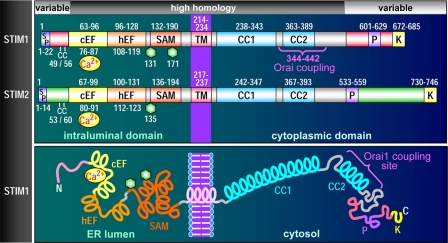
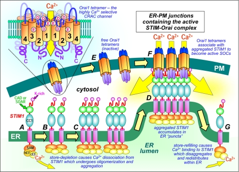
Ca(2+) signals controlling a vast array of cell functions involve both Ca(2+) store release and external Ca(2+) entry. These two events are coordinated through a dynamic intermembrane coupling between two distinct membrane proteins, STIM and Orai. STIM proteins are endoplasmic reticulum (ER) luminal Ca(2+) sensors that undergo a profound redistribution into discrete junctional ER domains closely juxtaposed with the plasma membrane (PM). Orai proteins are PM Ca(2+) channels that migrate and become tethered by STIM within the ER-PM junctions, where they mediate exceedingly selective Ca(2+) entry. We describe a new understanding of the nature of the proteins and how they function to mediate this remarkable intermembrane signaling process controlling Ca(2+) signals.


Figures

FIGURE 1.
FIGURE 1.
STIM protein domain structures. Upper, domain comparison of STIM1 and STIM2, including signal peptides (SP), a pair of highly conserved cysteines (CC), canonical cEF- and hEF-hands, SAMs, Asn-linked glycosylation sites (hexagons), TMs, coiled-coil regions (CC1 and CC2), proline-rich domains (P), and polybasic-rich domains (K). The minimal region of STIM1 known to be required for coupling to Orai1 is also shown (positions 344–442). Lower, diagrammatic representation of domain topology of STIM1.
FIGURE 2.
FIGURE 2.
Dynamic molecular coupling between STIM1 and Orai1 within ER-PM junctions. Depletion of ER luminal Ca2+ causes Ca2+ dissociation from the STIM1 N-terminal cEF-hand (A), leading to fast oligomerization of STIM1 due to unfolding and interactions between EF-SAM domains (B). Slower further aggregation of STIM1 through interactions between the C-terminal coiled-coil domain 1 (CC1) and CAD/SOAR domains (C) results in STIM1 diffusion, aggregation, and accumulation of STIM1 in pre-existing ER-PM junctions, stabilized by interactions of lysine-rich (K-rich) STIM1 N termini with the PM (D). Diffusible Orai1 tetramers in the PM (E) are trapped in junctions (F) by interaction with exposed CAD/SOAR domains, which bind to the C and N termini of Orai1 channels and conformationally gate the opening of Orai1 channels and Ca2+ entry. Upon store refilling, Ca2+ association with STIM1 reverses EF-SAM oligomerization, causing uncoupling from and deactivation of Orai1 and release of STIM1 monomers from puncta to redistribute around the ER (G). The topology of the Orai1 tetramer is shown (upper left), depicting the four transmembrane domains (1–4) and clusters of Asp (D) and Glu (E) residues that constitute the Ca2+-selective filter and pore.

Similar articles

Cited by

References

    1. Berridge M. J., Bootman M. D., Roderick H. L. (2003) Nat. Rev. Mol. Cell Biol. 4,517–529 - PubMed
    1. Zhang K., Kaufman R. J. (2008) Nature 454,455–462 - PMC - PubMed
    1. Parekh A. B., Penner R. (1997) Physiol. Rev. 77,901–930 - PubMed
    1. Venkatachalam K., van Rossum D. B., Patterson R. L., Ma H. T., Gill D. L. (2002) Nat. Cell Biol. 4,E263–E272 - PubMed
    1. Parekh A. B., Putney J. W., Jr. (2005) Physiol. Rev. 85,757–810 - PubMed

LinkOut - more resources