

Use of adipose stem cells and polylactide discs for tissue engineering of the temporomandibular joint disc
- PMID: 19474082
- PMCID: PMC2839381
- DOI: 10.1098/rsif.2009.0117
Use of adipose stem cells and polylactide discs for tissue engineering of the temporomandibular joint disc
Abstract
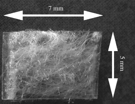
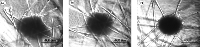
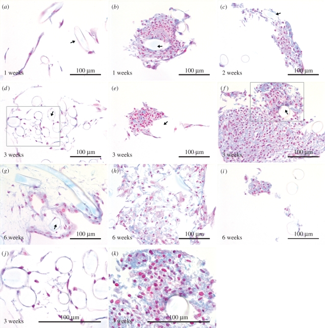
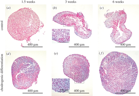
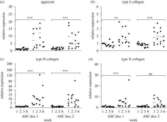
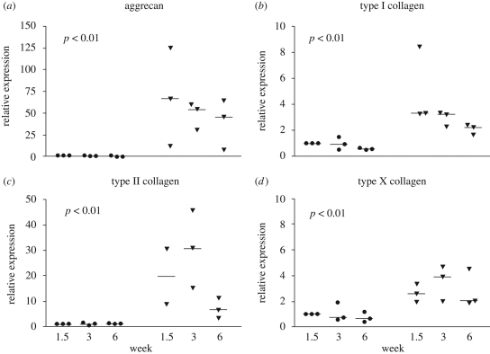
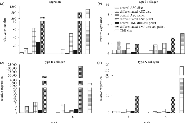
There is currently no suitable replacement for damaged temporomandibular joint (TMJ) discs after discectomy. In the present study, we fabricated bilayer biodegradable polylactide (PLA) discs comprising a non-woven mat of poly(L/D)lactide (P(L/D)LA) 96/4 and a P(L/DL)LA 70/30 membrane plate. The PLA disc was examined in combination with adipose stem cells (ASCs) for tissue engineering of the fibrocartilaginous TMJ disc in vitro. ASCs were cultured in parallel in control and chondrogenic medium for a maximum of six weeks. Relative expression of the genes, aggrecan, type I collagen and type II collagen present in the TMJ disc extracellular matrix increased in the ASC-seeded PLA discs in the chondrogenic medium. The hypertrophic marker, type X collagen, was moderately induced. Alcian blue staining showed accumulation of sulphated glycosaminoglycans. ASC differentiation in the PLA discs was close to that observed in pellet cultures. Comparison of the mRNA levels revealed that the degree of ASC differentiation was lower than that in TMJ disc-derived cells and tissue. The pellet format supported the phenotype of the TMJ disc-derived cells under chondrogenic conditions and also enhanced their hyalinization potential, which is considered part of the TMJ disc degeneration process. Accordingly, the combination of ASCs and PLA discs has potential for the development of a tissue-engineered TMJ disc replacement.
Figures
Similar articles
-
Autologous adipose stem cells and polylactide discs in the replacement of the rabbit temporomandibular joint disc.J R Soc Interface. 2013 May 29;10(85):20130287. doi: 10.1098/rsif.2013.0287. Print 2013 Aug 6. J R Soc Interface. 2013. PMID: 23720535 Free PMC article.
-
Fibro/chondrogenic differentiation of dental stem cells into chitosan/alginate scaffolds towards temporomandibular joint disc regeneration.J Mater Sci Mater Med. 2018 Jun 26;29(7):97. doi: 10.1007/s10856-018-6109-6. J Mater Sci Mater Med. 2018. PMID: 29946796
-
Effects of chitosan and bioactive glass modifications of knitted and rolled polylactide-based 96/4 L/D scaffolds on chondrogenic differentiation of adipose stem cells.J Tissue Eng Regen Med. 2015 Jan;9(1):55-65. doi: 10.1002/term.1614. Epub 2012 Oct 22. J Tissue Eng Regen Med. 2015. PMID: 23086809
-
[Cell sources for engineered temporomandibular joint disc tissue: present and future].Sheng Wu Yi Xue Gong Cheng Xue Za Zhi. 2010 Apr;27(2):463-6. Sheng Wu Yi Xue Gong Cheng Xue Za Zhi. 2010. PMID: 20481340 Review. Chinese.
-
Design characteristics for temporomandibular joint disc tissue engineering: learning from tendon and articular cartilage.Proc Inst Mech Eng H. 2007 Jul;221(5):509-26. doi: 10.1243/09544119JEIM158. Proc Inst Mech Eng H. 2007. PMID: 17822153 Review.
Cited by
-
Autologous adipose stem cells and polylactide discs in the replacement of the rabbit temporomandibular joint disc.J R Soc Interface. 2013 May 29;10(85):20130287. doi: 10.1098/rsif.2013.0287. Print 2013 Aug 6. J R Soc Interface. 2013. PMID: 23720535 Free PMC article.
-
An Update on Mesenchymal Stem Cell-Centered Therapies in Temporomandibular Joint Osteoarthritis.Stem Cells Int. 2021 Apr 1;2021:6619527. doi: 10.1155/2021/6619527. eCollection 2021. Stem Cells Int. 2021. PMID: 33868408 Free PMC article. Review.
-
Tissue Engineering for the Temporomandibular Joint.Adv Healthc Mater. 2019 Jan;8(2):e1801236. doi: 10.1002/adhm.201801236. Epub 2018 Dec 17. Adv Healthc Mater. 2019. PMID: 30556348 Free PMC article. Review.
-
Effects of Macromolecular Crowding on Human Adipose Stem Cell Culture in Fetal Bovine Serum, Human Serum, and Defined Xeno-Free/Serum-Free Conditions.Stem Cells Int. 2017;2017:6909163. doi: 10.1155/2017/6909163. Epub 2017 Mar 30. Stem Cells Int. 2017. PMID: 28465691 Free PMC article.
-
940 nm diode laser induced differentiation of human adipose derived stem cells to temporomandibular joint disc cells.BMC Biotechnol. 2022 Aug 29;22(1):23. doi: 10.1186/s12896-022-00754-6. BMC Biotechnol. 2022. PMID: 36038860 Free PMC article.
References
-
- Ali A. M., Sharawy M. 1996a. Histochemical and immunohistochemical studies of the effects of experimental anterior disc displacement on sulfated glycosaminoglycans, hyaluronic acid, and link protein of the rabbit craniomandibular joint. J. Oral Maxillofac. Surg. 54, 992–1004. (10.1016/S0278-2391(96)90399-7) - DOI - PubMed
Publication types
MeSH terms
Substances
LinkOut - more resources
Full Text Sources
Medical
Miscellaneous
