. 2009 Aug;83(15):7739-48.
doi: 10.1128/JVI.00614-09. Epub 2009 May 27.

Human bocavirus can be cultured in differentiated human airway epithelial cells

Affiliations

Human bocavirus can be cultured in differentiated human airway epithelial cells

Ronald Dijkman et al. J Virol. 2009 Aug.

Abstract

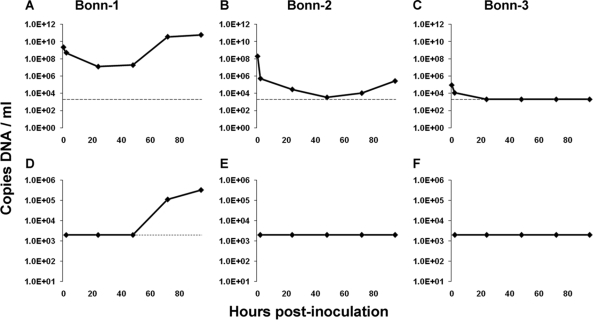
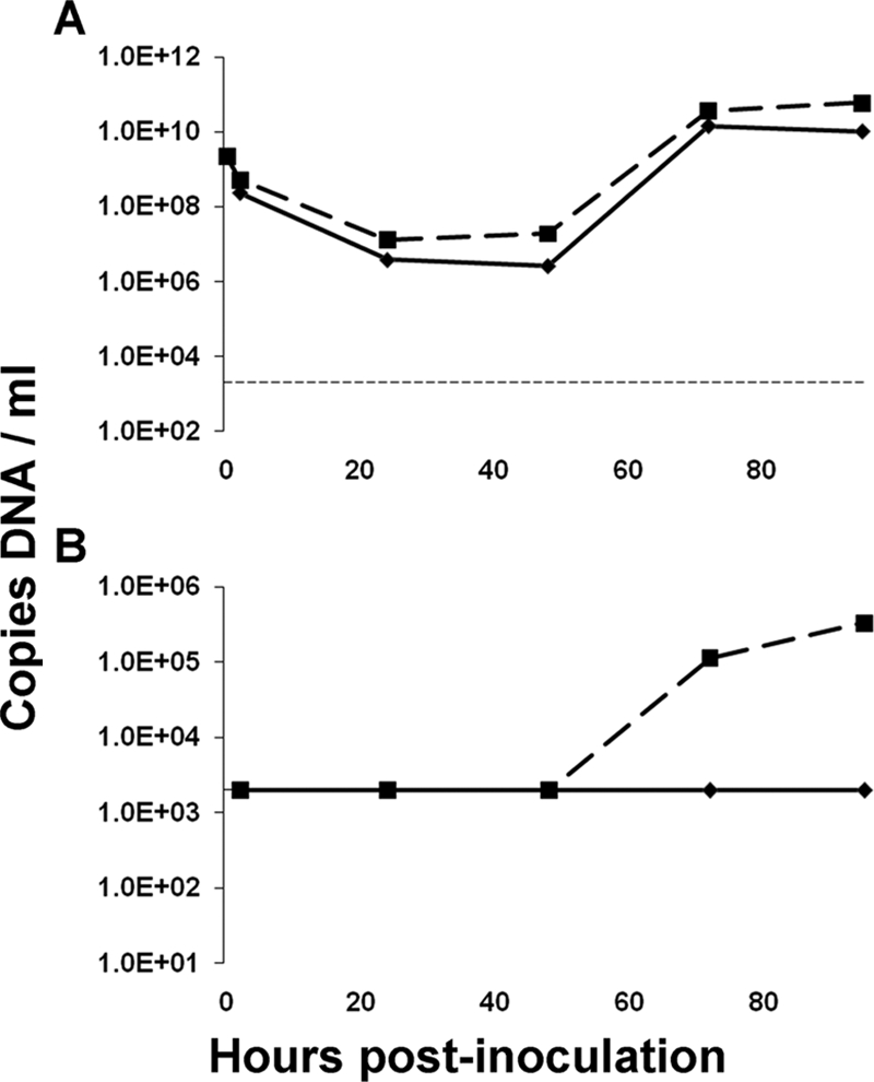
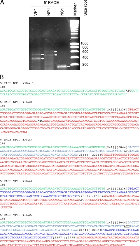
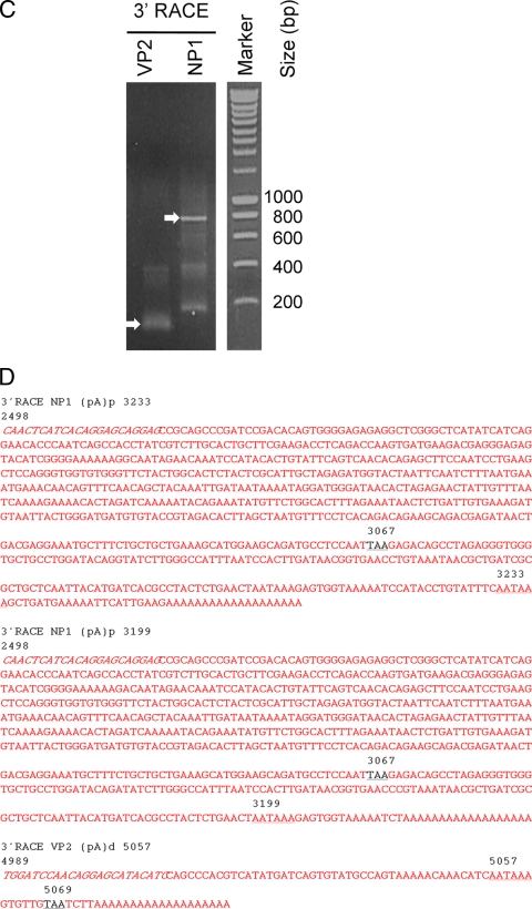
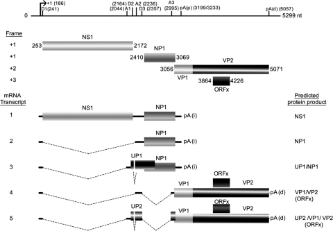
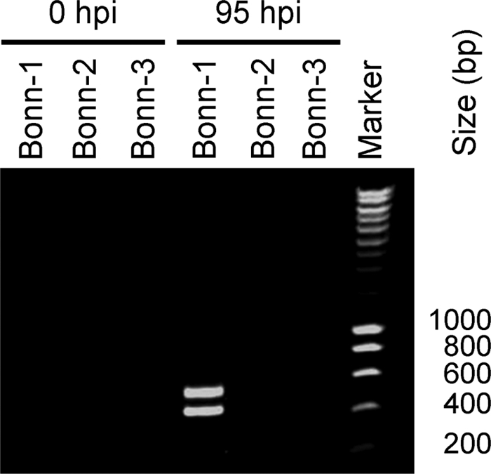
In 2005, a human bocavirus was discovered in children with respiratory tract illnesses. Attempts to culture this virus on conventional cell lines has failed thus far. We investigated whether the virus can replicate on pseudostratified human airway epithelium. This cell culture system mimics the human airway environment and facilitates culturing of various respiratory agents. The cells were inoculated with human bocavirus-positive nasopharyngeal washes from children, and virus replication was monitored by measuring apical release of the virus via real-time PCR. Furthermore, we identified different viral mRNAs in the infected cells. All mRNAs were transcribed from a single promoter but varied due to alternative splicing and alternative polyadenylation, similar to what has been described for bovine parvovirus and minute virus of canines, the other two members of the Bocavirus genus. Thus, transcription of human bocavirus displays strong homology to the transcription of the other bocaviruses. In conclusion, we report here for the first time that human bocavirus can be propagated in an in vitro culture system and present a detailed map of the set of mRNAs that are produced by the virus.


Figures

FIG. 1.
FIG. 1.
HBoV propagation on human airway epithelium cell culture. HBoV DNA concentration (copies of DNA/ml; y axis) in the apical washings at different hours postinoculation (x axis) for the Bonn-1 (A), Bonn-2 (B), and Bonn-3 (C) HBoV-inoculated human airway epithelium cell cultures. HBoV DNA quantification at the basolateral side is shown in panels D (Bonn-1), E (Bonn-2), and F (Bonn-3). The horizontal dashed line represents the detection threshold of the HBoV DNA real-time PCR assay.
FIG. 2.
FIG. 2.
DNase protection assay of HBoV DNA from the Bonn-1 culture. HBoV DNA concentration (copies of DNA/ml; y axis) in DNase-treated (continuous line) and non-DNase-treated (dashed line) apical harvests (A) and basolateral harvests (B). The horizontal dashed line in panel A represents the detection threshold of the assay.
FIG. 3.
FIG. 3.
Identification of mRNA transcripts from Bonn-1. (A) Agarose gel with ethidium bromide-stained 5′ RACE PCR products of NS1, NP1, and VP1 mRNAs of the Bonn-1 isolate at 95 hpi. The arrows indicate 5′ RACE-amplified PCR products. (B) The determined cDNA nucleotide sequences of the NS1, NP1, and VP1 5′ RACE products. The start position of each transcript is indicated at the beginning of the nucleotide sequence. The spectrum of color highlights the nucleotide sequences corresponding with different regions along the genome. The genome positions at the splice donor and acceptor junction sites are shown in parentheses. The first start codon triplets along each fragment are indicated in underlined black letters. In mRNA 1, the start codon is for NS1; for mRNA 2, the start codon is for NP1; for mRNA 3, the start codon is for UP1; for mRNA 4, the start codon is for VP1; and for mRNA5, the start codon is for UP2. The binding region of the reverse 5′ RACE primer is shown in italics. (C) Agarose gel with ethidium bromide-stained 3′ RACE products of the NP1 and VP2 mRNAs of the Bonn-1 isolate at 95 hpi. The arrows indicate 3′ RACE-amplified PCR products that were properly primed on the polyadenosine tails. The additional fragments were generated by nonspecific priming of the RT primer at polyadenosine stretches along the HBoV genome. (D) The determined cDNA nucleotide sequences of the VP2 and NP1 3′ RACE products. The forward 3′ RACE primer is shown in italics. The position of the stop codon of the NP1 and VP2 transcripts is indicated with underlined black letters, and the proximal polyadenylation [(pA)p] (NP1 3′RACE products) and distal pA [(pA)d] (VP2 3′RACE product) are underlined. The genome position of the stop codon and the pA sites are indicated above the sequence.
FIG. 3.
FIG. 3.
Identification of mRNA transcripts from Bonn-1. (A) Agarose gel with ethidium bromide-stained 5′ RACE PCR products of NS1, NP1, and VP1 mRNAs of the Bonn-1 isolate at 95 hpi. The arrows indicate 5′ RACE-amplified PCR products. (B) The determined cDNA nucleotide sequences of the NS1, NP1, and VP1 5′ RACE products. The start position of each transcript is indicated at the beginning of the nucleotide sequence. The spectrum of color highlights the nucleotide sequences corresponding with different regions along the genome. The genome positions at the splice donor and acceptor junction sites are shown in parentheses. The first start codon triplets along each fragment are indicated in underlined black letters. In mRNA 1, the start codon is for NS1; for mRNA 2, the start codon is for NP1; for mRNA 3, the start codon is for UP1; for mRNA 4, the start codon is for VP1; and for mRNA5, the start codon is for UP2. The binding region of the reverse 5′ RACE primer is shown in italics. (C) Agarose gel with ethidium bromide-stained 3′ RACE products of the NP1 and VP2 mRNAs of the Bonn-1 isolate at 95 hpi. The arrows indicate 3′ RACE-amplified PCR products that were properly primed on the polyadenosine tails. The additional fragments were generated by nonspecific priming of the RT primer at polyadenosine stretches along the HBoV genome. (D) The determined cDNA nucleotide sequences of the VP2 and NP1 3′ RACE products. The forward 3′ RACE primer is shown in italics. The position of the stop codon of the NP1 and VP2 transcripts is indicated with underlined black letters, and the proximal polyadenylation [(pA)p] (NP1 3′RACE products) and distal pA [(pA)d] (VP2 3′RACE product) are underlined. The genome position of the stop codon and the pA sites are indicated above the sequence.
FIG. 4.
FIG. 4.
Schematic representation of the genomic and transcriptional map of HBoV Bonn-1 isolate. The transcriptional start (arrow), the splice donor (D numbers) and acceptor (A numbers) sites, the proximal and distal polyadenylation signals [pA(p) and pA(d), respectively], and the predicted ORFs are positioned along the HBoV genome of the Bonn-1 isolate. In the lower panel, a schematic overview of the identified mRNA transcripts of Bonn-1 HBoV and their suggested protein products is presented.
FIG. 5.
FIG. 5.
Spliced mRNAs are not present in the inoculum. Agarose gel with ethidium bromide-stained RT-PCR products. Primers spanning the splice junctions in the NP1 mRNA were used for amplification (primers BOCA_5UTR1 and BOCA_R12; Table 1). At 95 hpi, the spliced mRNA RT-PCR is positive (mRNA 2, 445 bp; mRNA 3, 565 bp) for the Bonn-1 HBoV, whereas the inoculum did not contain HBoV mRNA (0 hpi).

References

    1. Allander, T., T. Jartti, S. Gupta, H. G. Niesters, P. Lehtinen, R. Osterback, T. Vuorinen, M. Waris, A. Bjerkner, A. Tiveljung-Lindell, B. G. van den Hoogen, T. Hyypia, and O. Ruuskanen. 2007. Human bocavirus and acute wheezing in children. Clin. Infect. Dis. 44904-910. - PMC - PubMed
    1. Allander, T., M. T. Tammi, M. Eriksson, A. Bjerkner, A. Tiveljung-Lindell, and B. Andersson. 2005. Cloning of a human parvovirus by molecular screening of respiratory tract samples. Proc. Natl. Acad. Sci. USA 10212891-12896. - PMC - PubMed
    1. Berns, K., and C. R. Parrish. 2007. Parvoviridae, p. 2437-2477. In D. M. Knipe, P. M. Howley, D. E. Griffin, R. A. Lamb, M. A. Martin, B. Roizman, and S. E. Straus (ed.), Fields virology, 5th ed. Lippincott Williams & Wilkins, Philadelphia, PA.
    1. Berns, K. I. 1990. Parvovirus replication. Microbiol. Rev. 54316-329. - PMC - PubMed
    1. Blom, N., J. Hansen, D. Blaas, and S. Brunak. 1996. Cleavage site analysis in picornaviral polyproteins: discovering cellular targets by neural networks. Protein Sci. 52203-2216. - PMC - PubMed

Publication types

Associated data

LinkOut - more resources