. 2009 Sep;90(Pt 9):2191-200.
doi: 10.1099/vir.0.012104-0. Epub 2009 May 27.

Drosophila A virus is an unusual RNA virus with a T=3 icosahedral core and permuted RNA-dependent RNA polymerase

Affiliations

Drosophila A virus is an unusual RNA virus with a T=3 icosahedral core and permuted RNA-dependent RNA polymerase

Rebecca L Ambrose et al. J Gen Virol. 2009 Sep.

Abstract

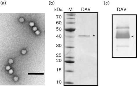
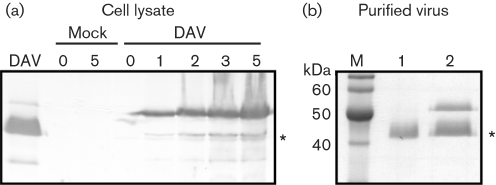
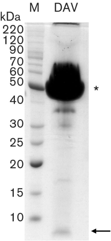
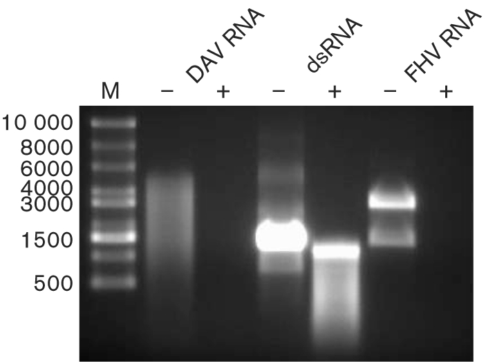
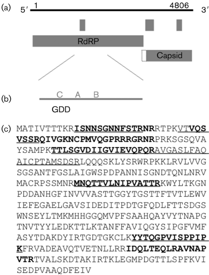
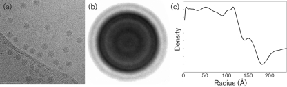
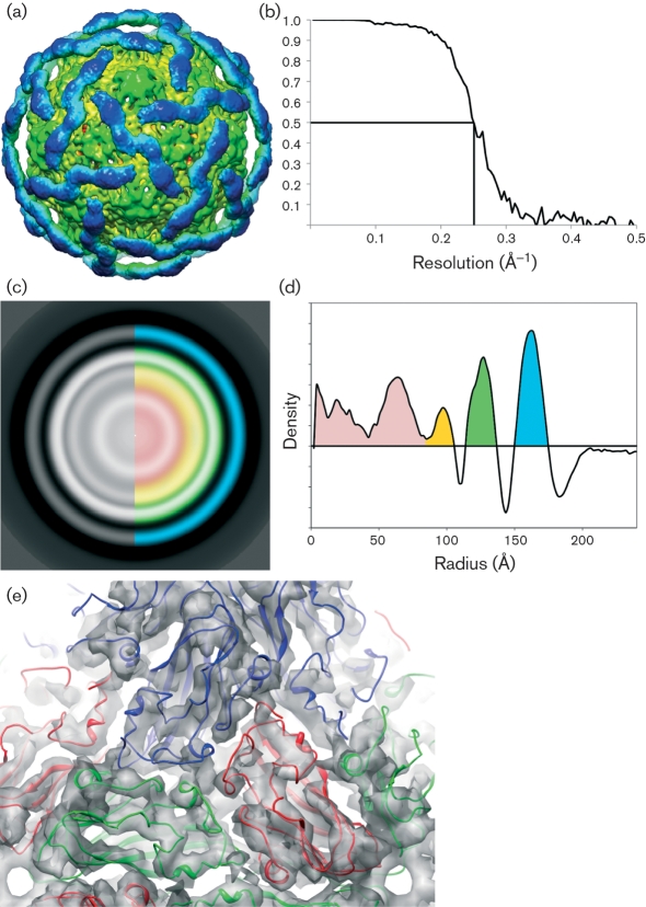
The vinegar fly, Drosophila melanogaster, is a popular model for the study of invertebrate antiviral immune responses. Several picorna-like viruses are commonly found in both wild and laboratory populations of D. melanogaster. The best-studied and most pathogenic of these is the dicistrovirus Drosophila C virus. Among the uncharacterized small RNA viruses of D. melanogaster, Drosophila A virus (DAV) is the least pathogenic. Historically, DAV has been labelled as a picorna-like virus based on its particle size and the content of its RNA genome. Here, we describe the characterization of both the genome and the virion structure of DAV. Unexpectedly, the DAV genome was shown to encode a circular permutation in the palm-domain motifs of the RNA-dependent RNA polymerase. This arrangement has only been described previously for a subset of viruses from the double-stranded RNA virus family Birnaviridae and the T=4 single-stranded RNA virus family Tetraviridae. The 8 A (0.8 nm) DAV virion structure computed from cryo-electron microscopy and image reconstruction indicates that the virus structural protein forms two discrete domains within the capsid. The inner domain is formed from a clear T=3 lattice with similarity to the beta-sandwich domain of tomato bushy stunt virus, whilst the outer domain is not ordered icosahedrally, but forms a cage-like structure that surrounds the core domain. Taken together, this indicates that DAV is highly divergent from previously described viruses.


Figures

Fig. 1.
Fig. 1.
Analysis of DAV virions and structural proteins. (a) Sucrose gradient-purified virions were negatively stained and visualized by TEM at ×100 000 magnification. Bar, 100 nm. (b) Proteins present in purified virus particles were separated by electrophoresis through a 4–12 % Bis–Tris gradient gel. Total protein was visualized by staining with Coomassie blue. The capsid protein is indicated by an asterisk. Sizes of molecular mass markers are shown on the left. (c) Virion proteins separated through an SDS–10 % polyacrylamide gel were transferred to a nitrocellulose membrane and detected with a rabbit polyclonal anti-DAV antibody. The band marked with an asterisk corresponds to the 42 kDa protein indicated in (b).
Fig. 2.
Fig. 2.
Detection of virus protein synthesis in infected Drosophila DL2 cells. (a) DL2 cells were either mock-infected or infected with DAV and incubated at 26 °C until harvesting. Cell samples were taken at 0, 1, 2, 3 and 5 days p.i. and lysed with sample buffer. Cellular proteins were separated by electrophoresis through an SDS–10 % polyacrylamide gel, transferred to a nitrocellulose membrane and incubated with an anti-DAV rabbit polyclonal antiserum to detect virus proteins. The asterisk indicates the 42 kDa capsid protein. Virus proteins were detected at 1 day p.i. and their levels increased over time. No DAV-specific bands were detected in the mock-infected samples. (b) Proteins from DAVHD virions purified from infected DL2 cells (lane 2) and adult Drosophila (lane 1) were electrophoresed through an SDS–10 % polyacrylamide gel stained with Coomassie blue. The asterisk indicates the 42 kDa capsid protein.
Fig. 3.
Fig. 3.
Identification of a small structural protein. Virion proteins were overloaded and separated by electrophoresis through an SDS–10 % polyacrylamide gel. Total protein was stained with Coomassie blue. The asterisk indicates the 42 kDa capsid protein band that is consistently observed in SDS-PAGE analyses. The arrow indicates the small protein band that is estimated to be approximately 6 kDa.
Fig. 4.
Fig. 4.
Analysis of DAV genomic nucleic acid. RNA was extracted from purified DAVHD virions and mock-treated (−) or treated with RNase ONE (Promega) (+), which degrades ssRNA only. As controls, a synthetic dsRNA product from DCV and the ssRNA genome from FHV were treated similarly. Following nuclease treatment, products were separated on a 1 % agarose gel and visualized by ethidium bromide staining. Sizes of the ssRNA markers (M; in nt) are indicated on the left.
Fig. 5.
Fig. 5.
Organization of the DAV genome. (a) The 4806 nt DAV genome encodes two major ORFs and three additional small ORFs that contain >50 aa. The ORFs as shown start with an AUG codon; the extension (dotted line) on the capsid ORF indicates that this ORF overlaps the RdRP ORF, but the first methionine is downstream of the RdRP ORF. (b) Analysis of the 5′-proximal ORF showed high similarity of DAV RdRP regions to those of members of the families Tetraviridae and Birnaviridae. These viruses have a permuted RdRP, with the GDD region upstream of other palm subdomains. DAV RdRP also showed this permuted arrangement, as indicated by C–A–B. (c) MS analysis indicates that the 3′-proximal large ORF encodes the capsid protein. The deduced sequence of the capsid protein is shown and the peptides identified by MALDI-TOF (in bold type) and LC-MS/MS (underlined) are indicated.
Fig. 6.
Fig. 6.
CryoEM analysis of DAV virions. (a) Representative electron micrograph of sucrose gradient-purified DAV in the frozen–hydrated state. (b) Averaged two-dimensional density corresponding to 22 000 centred raw particles. (c) One-dimensional radial plot of the averaged two-dimensional particles, showing discrete peaks for the capsid layers.
Fig. 7.
Fig. 7.
Analysis of three-dimensional reconstruction of DAV particles. (a) Surface representation of the DaV cryoEM reconstruction contoured at 2σ. The inner N-terminal domain (green) is well-ordered with visible secondary-structural features, whereas the outer C-terminal domain (blue) contains only very-low-resolution features. (b) Estimated resolution of the reconstruction is 8 Å (0.8 nm) according to FSC criteria (y-axis) at 0.5. (c) Radially averaged slice through the centre of reconstruction. Blue, C-terminal domain; green, N-terminal domain; orange and red, RNA. (d) Plot showing density by radius [colours as in (c)]. (e) Docking of the β-sandwich domain of the TBSV crystal structure into the N-terminal domain of the DaV cryoEM density.

References

    1. Agrawal, D. K. & Johnson, J. E. (1992). Sequence and analysis of the capsid protein of Nudaurelia capensis ω virus, an insect virus with T=4 icosahedral symmetry. Virology 190, 806–814. - PubMed
    1. Brun, G. & Plus, N. (1980). The viruses of Drosophila. In The Genetics and Biology of Drosophila, pp. 625–702. Edited by M. Ashburner & T. F. R. Wright. New York: Academic Press.
    1. Cherry, S. & Perrimon, N. (2004). Entry is a rate-limiting step for viral infection in a Drosophila melanogaster model of pathogenesis. Nat Immunol 5, 81–87. - PMC - PubMed
    1. Cherry, S., Doukas, T., Armknecht, S., Whelan, S., Wang, H., Sarnow, P. & Perrimon, N. (2005). Genome-wide RNAi screen reveals a specific sensitivity of IRES-containing RNA viruses to host translation inhibition. Genes Dev 19, 445–452. - PMC - PubMed
    1. Christian, P. D. (1992). A simple vacuum dot-blot hybridisation assay for the detection of Drosophila A and C viruses in single Drosophila. J Virol Methods 38, 153–165. - PubMed

Publication types

MeSH terms

Associated data

LinkOut - more resources