. 2009 May 27;29(21):6771-9.
doi: 10.1523/JNEUROSCI.0887-09.2009.

Human APOE isoform-dependent effects on brain beta-amyloid levels in PDAPP transgenic mice

Affiliations

Human APOE isoform-dependent effects on brain beta-amyloid levels in PDAPP transgenic mice

Kelly R Bales et al. J Neurosci. .

Abstract

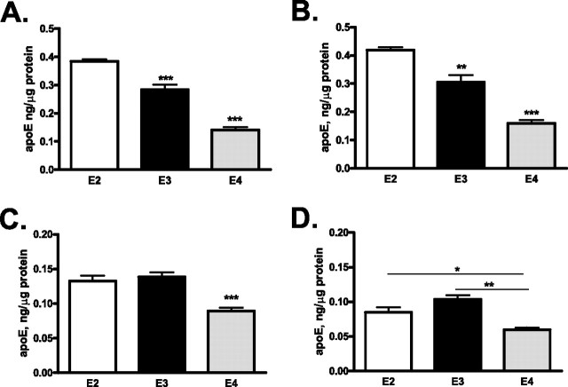
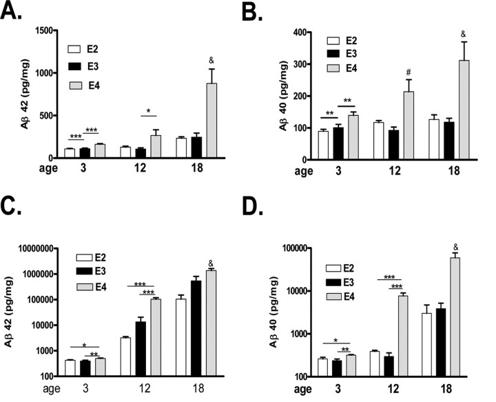
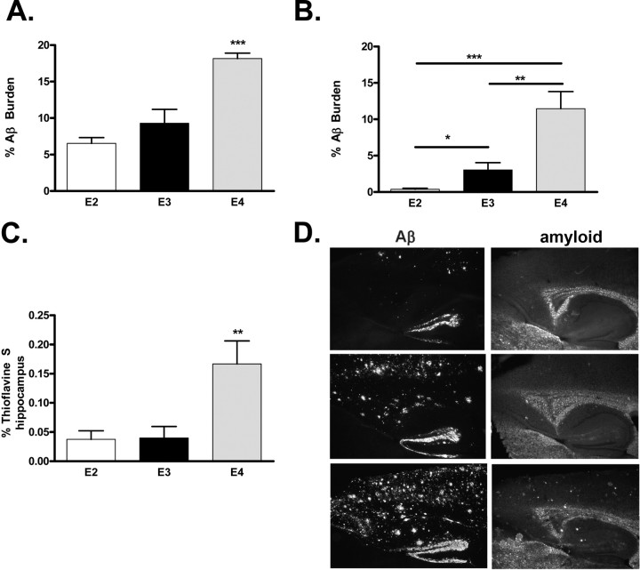
To investigate the role of human apolipoprotein E (apoE) on Abeta deposition in vivo, we crossed PDAPP mice lacking mouse Apoe to targeted replacement mice expressing human apoE (PDAPP/TRE2, PDAPP/TRE3, or PDAPP/TRE4). We then measured the levels of apoE protein and Abeta peptides in plasma, CSF, and brain homogenates in these mice at different ages. We also quantified the amount of brain Abeta and amyloid burden in 18-month-old mice. In young PDAPP/TRE4 mice that were analyzed at an age before brain Abeta deposition, we observed a significant decrease in the levels of apoE in CSF and brain when compared with age-matched mice expressing either human E2 or E3. The brain levels of Abeta42 in PDAPP/TRE4 mice were substantially elevated even at this very early time point. In older PDAPP/TRE4 mice, the levels of insoluble apoE protein increased in parallel to the dramatic rise in brain Abeta burden, and the majority of apoE was associated with Abeta. In TRE4 only mice, we also observed a significant decrease in the level of apoE in brain homogenates. Since the relative level of apoE mRNA was equivalent in PDAPP/TRE and TRE only mice, it appears that post-translational mechanisms influence the levels of apoE protein in brain (E4 < E3 << E2), resulting in early and dramatic apoE isoform-dependent effects on brain Abeta levels (E4 >> E3 > E2) that increase with age. Therapeutic strategies aimed at increasing the soluble levels of apoE protein, regardless of isoform, may effectively prevent and (or) treat Alzheimer's disease.


Figures

Figure 1.
Figure 1.
A–F, Levels of apoE in plasma, CSF, and brain homogenates in PDAPP mice expressing human apoE at various ages. A, B, ApoE levels in plasma (A) and CSF (B) were significantly greater in PDAPP/TRE2 mice, whereas the levels of apoE were the lowest in PDAPP/TRE4 mice. C, D, Soluble levels of apoE in hippocampal (C) or cortical (D) homogenates were significantly greater in PDAPP/TRE2 mice at all ages examined, whereas the levels of soluble apoE were significantly lower in PDAPP/TRE4 mice. E, F, Insoluble levels of apoE measured in the hippocampus (E) were significantly lower in PDAPP/TRE4 mice at a young age. There was an age-dependent increase in insoluble apoE levels in hippocampus (E) and cortex (F) of PDAPP mice expressing one of the human apoE alleles. A, ***p < 0.001 versus all other groups ANOVA, Tukey–Kramer post test, *p < 0.05 t test. B, ***p < 0.001 versus all other groups ANOVA, Tukey–Kramer post test, **p < 0.01 t test. C, ∧∧∧ p < 0.001 versus all other groups, ### p < 0.001 versus all other groups except # p < 0.05 versus E4 at 3 months ANOVA, Tukey–Kramer post test, *p < 0.05, **p < 0.01, ***p < 0.001 t test. D, ∧∧∧ p < 0.001 versus all other groups, ### p < 0.001 versus all other groups except E4 at 3 months, & p < 0.001 versus all other groups except E2 and E3 at 3 months ANOVA, Tukey–Kramer post test, **p < 0.05, ***p < 0.001 t test. E, & p < 0.001 versus all other groups except E4 at 18 months p < 0.05 versus E3 at 12 months, # p < 0.05 versus E4 at 18 months, ANOVA, Tukey–Kramer post test, *p < 0.05 t test. F, & p < 0.001 versus all other groups, # p < 0.05 versus al other groups, ANOVA, Tukey–Kramer post test.
Figure 2.
Figure 2.
A–D, Levels of apoE in brain homogenates from mice expressing human apoE2, 3, or 4 (E2, E3, or E4; 3 months of age) only. A, B, Soluble levels of apoE in hippocampal (A) or cortical (B) homogenates were significantly greater in TRE2 mice, whereas the levels of soluble apoE were significantly lower in TRE4 mice. C, D, Insoluble levels of apoE measured in the hippocampus (C) or cortex (D) were significantly lower in TRE4 mice. A–C, ***p < 0.001, **p < 0.01 versus all other groups ANOVA, Tukey–Kramer post test. D, **p < 0.05, *p < 0.01 t test.
Figure 3.
Figure 3.
A–D, Levels of Aβ in plasma (A, B) or CSF (C, D) in PDAPP mice expressing one of the human apolipoprotein E alleles (E2, E3, or E4) at 3 or 18 months. B, C, ***p < 0.001 versus all groups at 18 months of age, **p < 0.01 versus E2 at 3 months of age, *p < 0.01 versus E4 at 3 months of age, ANOVA, Tukey–Kramer post test, ***p < 0.001, **p < 0.01 t test.
Figure 4.
Figure 4.
A–D, Levels of soluble (A, B) and insoluble (C, D) Aβ in hippocampal homogenates from PDAPP mice expressing human apolipoprotein E alleles at different ages. A, & p < 0.001 versus all groups, ### p < 0.001 versus E2 or E3 at 12 months; E4 at 3 months, # p < 0.01 versus E3 18 months, ANOVA, Tukey–Kramer post test, ***p < 0.001, t test. B, & p < 0.001 versus all groups except E4 at 12 months, # p < 0.01 versus E4 at 3 months and E3 at 12 months, ANOVA, Tukey–Kramer post test, ***p < 0.001 t test. C, & p < 0.01 versus all other groups expect E3 at 18 months; # p < 0.01 versus all other groups except E4 at 18 months; ∧p < 0.01 versus E2 at 3 and 12 months, ANOVA, Tukey–Kramer post test, **p < 0.01, ***p < 0.001, t test. D, & p < 0.001 versus all other groups, ANOVA, Tukey–Kramer post test, ***p < 0.001, **p < 0.05 t test.
Figure 5.
Figure 5.
A–D, Levels of soluble (A, B) and insoluble Aβ (C, D) in cortical homogenates from PDAPP mice expressing human apolipoprotein E alleles at different ages. A, & p < 0.001 versus all other groups, ANOVA, Tukey–Kramer post test, *p < 0.05, ***p < 0.001 t test. B, & p < 0.001 versus all other groups except E4 at 12 months, # p < 0.05 versus E3 at 12 months ANOVA, Tukey–Kramer post test, **p < 0.01, t test. C, & p < 0.001 versus all other groups, ANOVA, Tukey–Kramer post test, *p < 0.05, **p < 0.01, ***p < 0.001, t test. D, & p < 0.001 versus all other groups, ANOVA, Tukey–Kramer post test, *p < 0.05, **p < 0.01, ***p < 0.001, t test.
Figure 6.
Figure 6.
A–D, Brain Aβ and amyloid burden in PDAPP mice expressing human apolipoprotein E alleles at 18 months of age. Significantly more Aβ is deposited in the hippocampus (A, ***p < 0.001, ANOVA, Tukey–Kramer post test) or cortex (B, ***p < 0.001, **p < 0.01, *p < 0.05, t test) of PDAPP/TRE4 mice at 18 months of age. Significantly more amyloid (thioflavine S material) is present in the hippocampus of PDAPP/TRE4 mice at 18 months of age (C, **p < 0.01, ANOVA, Tukey–Kramer post test). D, Representative photomicrographs of Aβ immunoreactivity and amyloid (thioflavine S staining) in PDAPP mice expressing various human apoE alleles at 18 months of age.
Figure 7.
Figure 7.
A, Representative images from 18-month-old PDAPP/TRE mice double stained with anti-apoE (apoE, green) or anti-Aβ (Aβ, red) and merged (yellow). B, Quantification of apoE and Aβ colocalization in PDAPP/TRE brain sections. The vast majority (∼90%) the apoE signal that is measured in the brain from PDAPP/TRE4 mice is associated with Aβ, whereas only ∼25% of apoE and Aβ are colocalized in very old PDAPP/TRE2 mice. ***p < 0.001 versus all other groups, ANOVA, Tukey–Kramer post test.
Figure 8.
Figure 8.
Significant inverse correlation between soluble levels of apolipoprotein E2 and Aβ 42 extracted from the hippocampus of PDAPP mice expressing human E2 (r 2 = 0.46, p < 0.001).

References

    1. Bales KR, Verina T, Dodel RC, Du Y, Altstiel L, Bender M, Hyslop P, Johnstone EM, Little SP, Cummins DJ, Piccardo P, Ghetti B, Paul SM. Lack of apolipoprotein E dramatically reduces amyloid β-peptide deposition. Nat Genet. 1997;17:263–264. - PubMed
    1. Bales KR, Verina T, Cummins DJ, Du Y, Dodel RC, Saura J, Fishman CE, DeLong CA, Piccardo P, Petegnief V, Ghetti B, Paul SM. Apolipoprotein E is essential for amyloid deposition in the APP V717F transgenic mouse model of Alzheimer's disease. Proc Natl Acad Sci U S A. 1999;96:15233–15238. - PMC - PubMed
    1. Beffert U, Cohn JS, Petit-Turcotte C, Tremblay M, Aumont N, Ramassamy C, Davignon J, Poirier J. Apolipoprotein E and beta-amyloid levels in the hippocampus and frontal cortex of Alzheimer's disease subjects are disease-related and apolipoprotein E genotype dependent. Brain Res. 1999;843:87–94. - PubMed
    1. Boschert U, Merlo-Pich E, Higgins G, Roses AD, Catsicas S. Apolipoprotein E expression by neurons surviving excitotoxic stress. Neurobiol Dis. 1999;6:508–514. - PubMed
    1. Boyles JK, Pitas RE, Wilson E, Mahley RW, Taylor JM. Apolipoprotein E associated with astrocytic glia of the central nervous system and with nonmylinating glia of the peripheral nervous system. J Clin Invest. 1985;76:1501–1513. - PMC - PubMed

LinkOut - more resources