

Stability of thalamocortical synaptic transmission across awake brain states
- PMID: 19474312
- PMCID: PMC2713605
- DOI: 10.1523/JNEUROSCI.5983-08.2009
Stability of thalamocortical synaptic transmission across awake brain states
Abstract
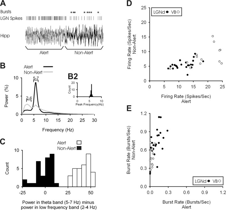
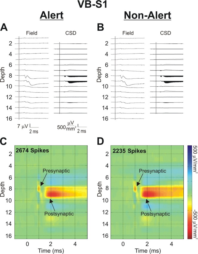
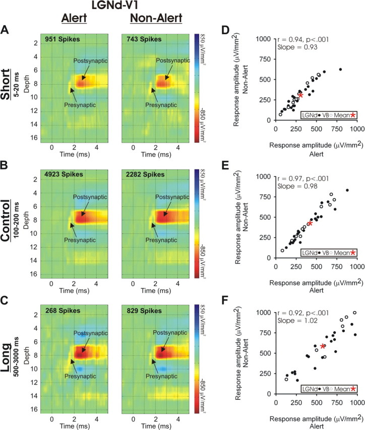
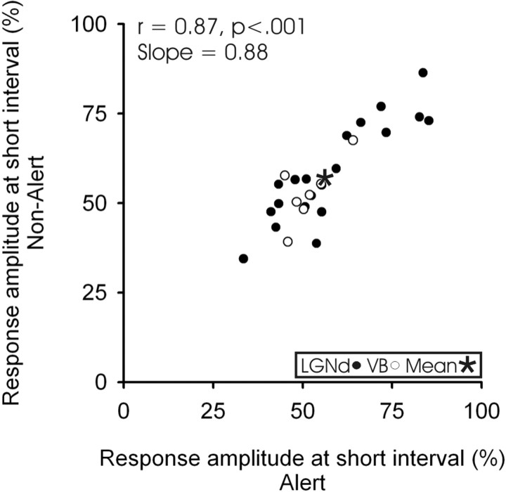
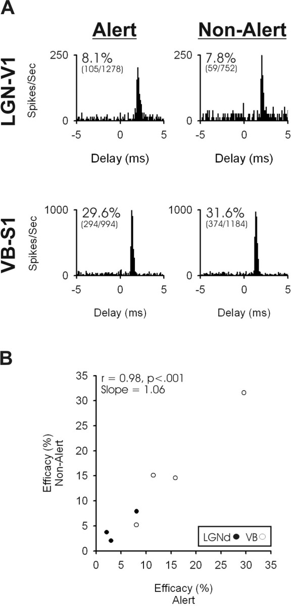
Sensory cortical neurons are highly sensitive to brain state, with many neurons showing changes in spatial and/or temporal response properties and some neurons becoming virtually unresponsive when subjects are not alert. Although some of these changes are undoubtedly attributable to state-related filtering at the thalamic level, another likely source of such effects is the thalamocortical (TC) synapse, where activation of nicotinic receptors on TC terminals have been shown to enhance synaptic transmission in vitro. However, monosynaptic TC synaptic transmission has not been directly examined during different states of alertness. Here, in awake rabbits that shifted between alert and non-alert EEG states, we examined the monosynaptic TC responses and short-term synaptic dynamics generated by spontaneous impulses of single visual and somatosensory TC neurons. We did this using spike-triggered current source-density analysis, an approach that enables assessment of monosynaptic extracellular currents generated in different cortical layers by impulses of single TC afferents. Spontaneous firing rates of TC neurons were higher, and burst rates were much lower in the alert state. However, we found no state-related changes in the amplitude of monosynaptic TC responses when TC spikes with similar preceding interspike interval were compared. Moreover, the relationship between the preceding interspike interval of the TC spike and postsynaptic response amplitude was not influenced by state. These data indicate that TC synaptic transmission and dynamics are highly conserved across different states of alertness and that observed state-related changes in receptive field properties that occur at the cortical level result from other mechanisms.
Figures
Similar articles
-
Spike timing and synaptic dynamics at the awake thalamocortical synapse.Prog Brain Res. 2005;149:91-105. doi: 10.1016/S0079-6123(05)49008-1. Prog Brain Res. 2005. PMID: 16226579 Review.
-
Activation of a cortical column by a thalamocortical impulse.J Neurosci. 2002 Sep 1;22(17):7766-73. doi: 10.1523/JNEUROSCI.22-17-07766.2002. J Neurosci. 2002. PMID: 12196600 Free PMC article.
-
The impact of an LGNd impulse on the awake visual cortex: synaptic dynamics and the sustained/transient distinction.J Neurosci. 2008 May 7;28(19):5018-28. doi: 10.1523/JNEUROSCI.4726-07.2008. J Neurosci. 2008. PMID: 18463255 Free PMC article.
-
Focal Local Field Potential Signature of the Single-Axon Monosynaptic Thalamocortical Connection.J Neurosci. 2017 May 17;37(20):5123-5143. doi: 10.1523/JNEUROSCI.2715-16.2017. Epub 2017 Apr 21. J Neurosci. 2017. PMID: 28432143 Free PMC article.
-
Thalamocortical control of feed-forward inhibition in awake somatosensory 'barrel' cortex.Philos Trans R Soc Lond B Biol Sci. 2002 Dec 29;357(1428):1717-27. doi: 10.1098/rstb.2002.1156. Philos Trans R Soc Lond B Biol Sci. 2002. PMID: 12626006 Free PMC article. Review.
Cited by
-
Predictable Fluctuations in Excitatory Synaptic Strength Due to Natural Variation in Presynaptic Firing Rate.J Neurosci. 2022 Nov 16;42(46):8608-8620. doi: 10.1523/JNEUROSCI.0808-22.2022. Epub 2022 Sep 28. J Neurosci. 2022. PMID: 36171085 Free PMC article.
-
Thalamic state influences timing precision in the thalamocortical circuit.J Neurophysiol. 2021 May 1;125(5):1833-1850. doi: 10.1152/jn.00261.2020. Epub 2021 Mar 24. J Neurophysiol. 2021. PMID: 33760642 Free PMC article.
-
A novel method for classifying cortical state to identify the accompanying changes in cerebral hemodynamics.J Neurosci Methods. 2016 Jul 15;267:21-34. doi: 10.1016/j.jneumeth.2016.04.005. Epub 2016 Apr 7. J Neurosci Methods. 2016. PMID: 27063501 Free PMC article.
-
Extracting functional components of neural dynamics with Independent Component Analysis and inverse Current Source Density.J Comput Neurosci. 2010 Dec;29(3):459-73. doi: 10.1007/s10827-009-0203-1. Epub 2009 Dec 22. J Comput Neurosci. 2010. PMID: 20033271
-
Layer-Specific Physiological Features and Interlaminar Interactions in the Primary Visual Cortex of the Mouse.Neuron. 2019 Feb 6;101(3):500-513.e5. doi: 10.1016/j.neuron.2018.12.009. Epub 2019 Jan 8. Neuron. 2019. PMID: 30635232 Free PMC article.
References
-
- Aston-Jones G. Brain structures and receptors involved in alertness. Sleep Med. 2005;6(Suppl 1):S3–S7. - PubMed
-
- Bacci A, Huguenard JR, Prince DA. Modulation of neocortical interneurons: extrinsic influences and exercises in self-control. Trends Neurosci. 2005;28:602–610. - PubMed
-
- Bartlett JR, Doty RW, Pecci-Saavedra J, Wilson PD. Mesencephalic control of lateral geniculate nucleus in primates 3. Modifications with state of alertness. Exp Brain Res. 1973;18:214–224. - PubMed
-
- Bezdudnaya T, Cano M, Swadlow HA, Alonso JM. Temporal frequency tuning of excitatory and inhibitory neurons in alert and drowsy visual cortex. Soc Neurosc Abstr. 2004;30:410–5.
Publication types
MeSH terms
Grants and funding
LinkOut - more resources
Full Text Sources