. 2009 May 27;29(21):6860-70.
doi: 10.1523/JNEUROSCI.0232-09.2009.

Akt signals through the mammalian target of rapamycin pathway to regulate CNS myelination

Affiliations

Akt signals through the mammalian target of rapamycin pathway to regulate CNS myelination

S Priyadarshini Narayanan et al. J Neurosci. .

Abstract

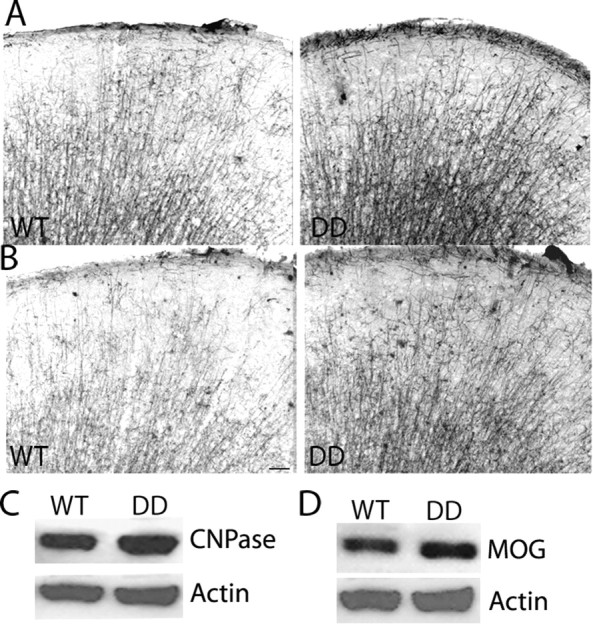
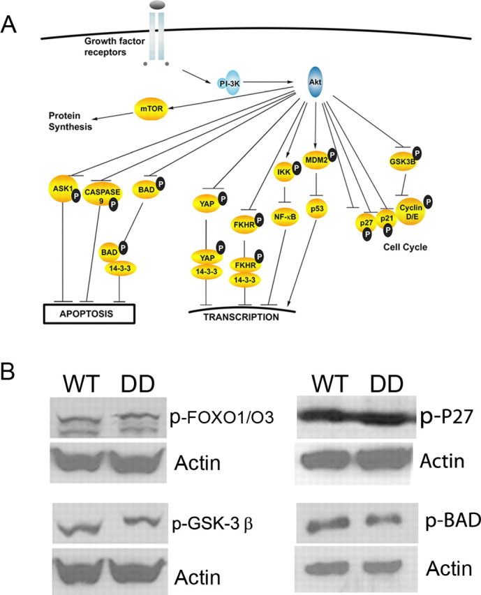
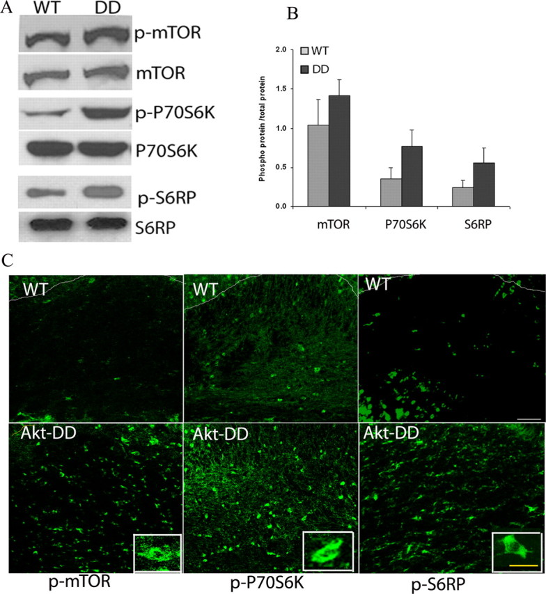
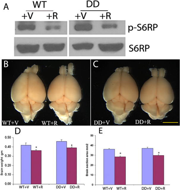
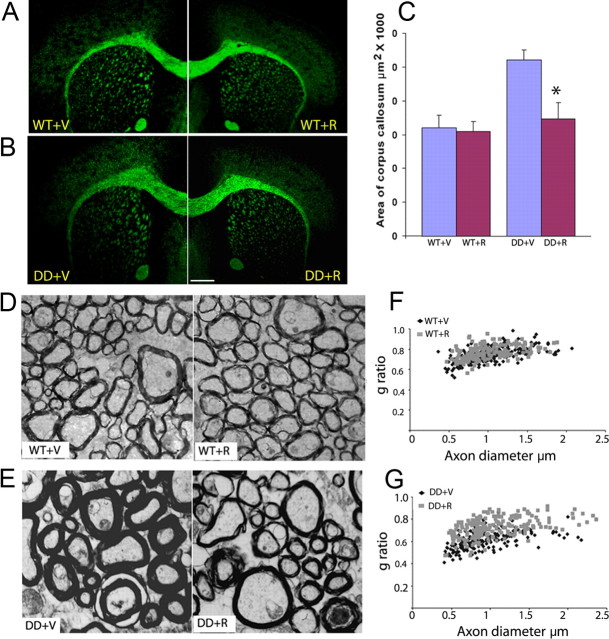
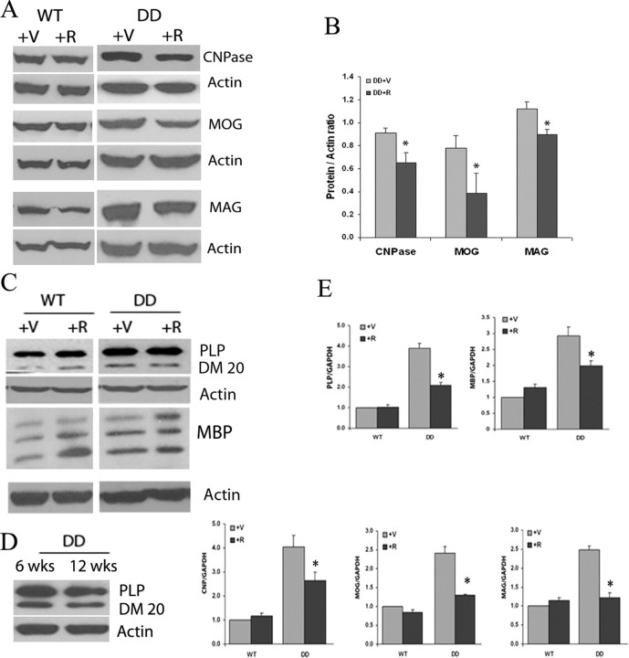
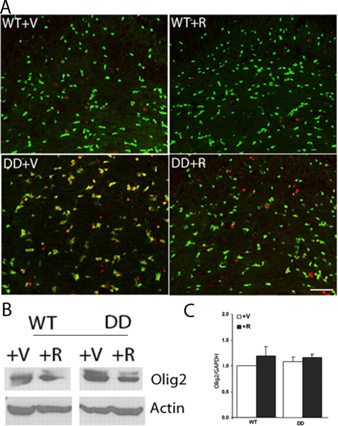
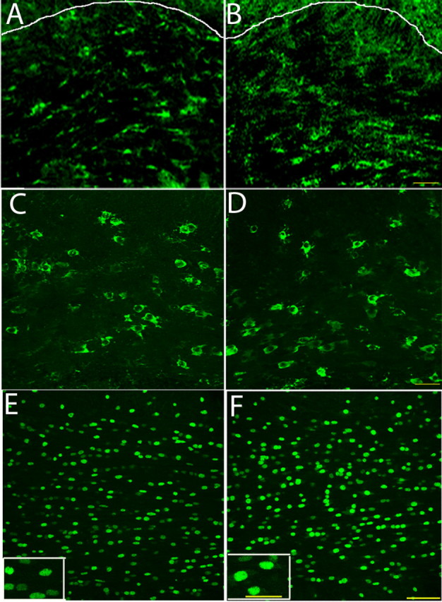
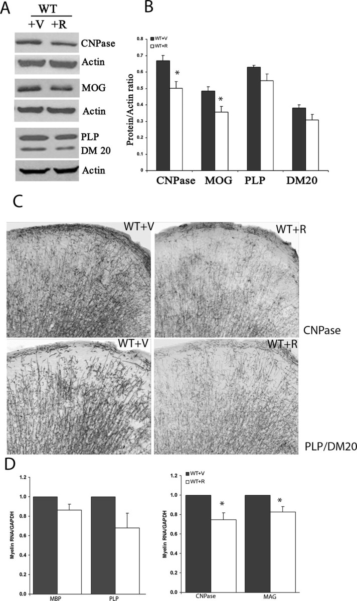
Mammalian target of rapamycin (mTOR), a well known Akt substrate, regulates multiple cellular functions including cell growth and protein synthesis. The current study identifies a novel role of the Akt/mTOR pathway as a regulator of CNS myelination. Previously, we showed that overexpressing constitutively active Akt in oligodendrocytes in a transgenic mouse model induces enhanced CNS myelination, with no changes in the proliferation or survival of oligodendrocyte progenitor or mature cells. The present study focused on the signaling mechanisms regulating this hypermyelination induced by Akt. Activation of mTOR and its downstream substrates (p70S6 kinase and S6 ribosomal protein) was observed in Akt-overexpressing oligodendrocytes. When mTOR signaling was inhibited chronically in vivo with rapamycin starting at 6 weeks of age, the observed hypermyelination was reduced to approximately the amount of myelin seen in wild-type mice. mTOR inhibition had little impact on wild-type myelination between 6 and 12 weeks of age, suggesting that, in normal adults, myelination is relatively complete and is no longer regulated by mTOR signaling. However, when mTOR was chronically inhibited in young adult wild-type mice, myelination was reduced. These results suggest that, during active myelination, the major Akt signal regulating CNS myelination is the mTOR pathway.


Figures

Figure 1.
Figure 1.
Enhanced myelination in Plp-Akt-DD mice. A, B, Coronal sections of cerebrum from 2-month-old WT and Plp-Akt-DD mice stained with antibodies against myelin proteins CNPase (A) and MOG (B). Note enhanced expression of both myelin proteins in Plp-Akt-DD brain relative to WT brain. A minimum of three animals were analyzed per genotype, and representative images are shown. Scale bar, 100 μm. C, D, Western blot data showing greater expression of CNPase (C) and MOG (D) in Plp-Akt-DD mice compared with WT mice at 2 months of age. CNPase expression relative to actin was 1.20 ± 0.08 (Plp-Akt-DD) compared with 0.97 ± 0.04 (WT); MOG expression relative to actin is 1.01 ± 0.11 (Plp-Akt-DD) compared with 0.71 ± 0.05 (WT). N = 3 and p < 0.05 in both cases.
Figure 2.
Figure 2.
Phosphorylation status of Akt substrates in Plp-Akt-DD brain. A, Schematic diagram indicating downstream substrates of Akt that regulate multiple cellular functions. B, Western blot analyses of cerebrum lysates from 2-month-old WT and Plp-Akt-DD mice for the Akt substrates GSK-3β, FOXO1/O3, p27, and BAD showed no differences in their phosphorylation status in Plp-Akt-DD samples, relative to WT. A minimum of three animals were analyzed per genotype, and representative blots are shown.
Figure 3.
Figure 3.
Activation of the mTOR pathway in Plp-Akt-DD brain. A, Representative Western blots showing the activation of mTOR, p70S6K, and S6RP in cerebrum lysates of 2-month-old Plp-Akt-DD samples and WT samples. B, Quantification of the amount of phosphorylated mTOR, p70S6K, and S6RP relative to the expression of their respective total protein levels. Note the increased phosphorylation of these proteins in Plp-Akt-DD mice compared with WT mice. N varies from 3 to 5. Error bars represent SEM. C, Immunohistochemistry of p-mTOR, p-p70S6K, and p-S6RP in 2-month-old WT and Plp-Akt-DD corpus callosum. Increased expression of these phosphorylated proteins in oligodendrocytes in Plp-Akt-DD corpus callosum confirmed that the activation of mTOR signaling in Plp-Akt-DD samples resulted from increased expression in oligodendrocytes. A minimum of three animals were analyzed per condition, and representative data are shown. The upper edge of the corpus callosum is outlined for WT samples (thin white line), but the corpus callosum fully fills the field in Plp-Akt-DD samples. Scale bar, 50 μm. The insets show oligodendrocytes immunostained for respective phosphoproteins, taken at high magnification. Scale bar, 20 μm.
Figure 4.
Figure 4.
Chronic rapamycin treatment impacted both WT and Plp-Akt-DD brains. Littermate WT and Plp-Akt-DD mice at 6–8 weeks of age were treated with rapamycin for 6 weeks. A, Representative Western blot showing decreased phosphorylation of S6RP in the brain lysates of rapamycin-treated WT and Plp-Akt-DD mice, compared with vehicle-treated WT and Plp-Akt-DD mice. N varies from 5 to 7 per condition. B, C, Representative images of WT (B) and Plp-Akt-DD (C) (12 weeks of age) brains after treatment with vehicle or rapamycin for 6 weeks. Relative to vehicle-treated animals, a reduction in the brain size of rapamycin-treated animals was noted for both WT and Plp-Akt-DD mice. N varies from 9 to 12 per condition. Scale bar, 5 mm. D, Quantification of the reduction in brain weight in rapamycin-treated WT and Plp-Akt-DD mice, compared with vehicle-treated animals. N varies from 9 to 12 per condition. *p < 0.005. E, Quantification of the reduction in brain sectional area for both rapamycin-treated WT and Plp-Akt-DD mice. Three consecutive sections from three different animals per condition were used for quantification. *p < 0.005. Error bars represent SEM.
Figure 5.
Figure 5.
Rapamycin treatment reduced the enhanced myelination in Plp-Akt-DD brains. Littermate WT and Plp-Akt-DD mice at 6–8 weeks of age were treated with rapamycin for 6 weeks. A, B, Confocal images showing the size of the corpus callosum in rapamycin-treated (R) Plp-Akt-DD sections (DD; bottom) or WT sections (WT; top), relative to vehicle-treated sections (V). Scale bar, 500 μm. C, Quantification of corpus callosum area, showing a significant reduction in rapamycin-treated Plp-Akt-DD animals, compared with the vehicle-treated group. N = 3; *p < 0.001. The small reduction in corpus callosum area in the WT group, however, was not significant. D, Electron micrograph images of optic nerves from vehicle-treated and rapamycin-treated WT mice, showing no obvious differences in the thickness of myelin after treatment. N = 3. Magnification, 10,000×. E, Electron micrographs of optic nerves from vehicle-treated and rapamycin-treated Plp-Akt-DD mice, showing a reduction of the enhanced myelination after rapamycin treatment. N = 3. Magnification, 10,000×. F, G, Quantification of the g ratios from myelinated axons in the rapamycin-treated and vehicle-treated groups of WT and Plp-Akt-DD optic nerves. In rapamycin-treated Plp-Akt-DD optic nerves, the g ratio was significantly increased, confirming the reduction in myelin thickness. N = 3; *p < 0.005. However, there was only a slight increase in the g ratio in rapamycin-treated WT optic nerve, compared with vehicle-treated samples. Error bars represent SEM.
Figure 6.
Figure 6.
Inhibition of mTOR signaling resulted in changes in the expression of myelin proteins and RNA levels in Plp-Akt-DD brains. Littermate WT and Plp-Akt-DD mice at 6–8 weeks of age were treated with rapamycin for 6 weeks. A, Western blot analysis showing the expression of the myelin proteins CNPase, MOG, and MAG in the rapamycin-treated WT and Plp-Akt-DD brains, compared with vehicle-treated controls for each group. N = 3 per condition, and representative data are shown. B, Quantification of Western blots showing a significant reduction in CNPase, MOG, and MAG in the brains of rapamycin-treated Plp-Akt-DD mice. N = 3; *p < 0.05. C, Western blot data showing no apparent change in the expression levels of the major myelin proteins PLP, DM20, and different MBP isoforms in the rapamycin-treated WT and Plp-Akt-DD samples, relative to vehicle-treated samples for each group. N = 3. D, Western blot analysis showing that the expression of the major myelin proteins PLP and DM20 does not detectably increase between the ages of 6 and 12 weeks in Plp-Akt-DD mice. E, Quantification of myelin RNA levels (CNPase, MOG, MAG, PLP, and MBP) using real-time PCR from the cerebrum samples of rapamycin-treated and vehicle-treated WT and Plp-Akt-DD brains. All myelin RNAs studied were downregulated in rapamycin-treated Plp-Akt-DD samples, compared with vehicle-treated controls. However, there were no significant changes in the levels of myelin RNAs in the rapamycin-treated WT samples. Quantification was performed relative to a standard RNA, GAPDH, and then expressed relative to the level of vehicle-treated WT sample, which was given an arbitrary value of 1.0. N = 3 per condition. *p < 0.05. Error bars represent SEM.
Figure 7.
Figure 7.
Expression of Olig2 is reduced in rapamycin-treated Plp-Akt-DD brains. Littermate WT and Plp-Akt-DD mice at 6–8 weeks of age were treated with rapamycin for 6 weeks. A, Immunohistochemistry showing Olig2 expression (red) in the corpus callosum of vehicle-treated and rapamycin-treated WT (WT; top) and Plp-Akt-DD (DD; bottom) coronal brain sections. Plp-Akt-DD and WT were crossed with Plp-EGFP mice to optimize visualization of green oligodendrocyte cell bodies. Olig2 expression was noted in Plp-Akt-DD oligodendrocytes but was undetectable in WT oligodendrocytes at this age. Olig2 was downregulated in rapamycin-treated Plp-Akt-DD oligodendrocytes. Scale bar, 50 μm. B, Western blot analysis using cerebrum lysates from vehicle-treated and rapamycin-treated WT and Plp-Akt-DD samples, showing reduced expression of Olig2 in the rapamycin-treated Plp-Akt-DD mice, compared with vehicle-treated Plp-Akt-DD controls. C, Quantitative real-time PCR analysis showing no changes in the expression of Olig2 RNA in rapamycin-treated Plp-Akt-DD or WT compared with vehicle-treated controls. Quantification was performed relative to a standard RNA, GAPDH, and then expressed relative to the level of vehicle-treated WT sample, which was given an arbitrary value of 1.0. N = 3 per condition. Error bars represent SEM.
Figure 8.
Figure 8.
Phospho-mTOR and Olig2 expression in WT and Plp-Akt-DD brains at P21. A, B, Immunohistochemistry of WT (A) and Plp-Akt-DD (B) corpus callosum analyzing p-mTOR expression showing the activation of mTOR in WT oligodendrocytes at P21. The upper edge of the corpus callosum is outlined for both WT and Plp-Akt-DD samples (thin white line). The expression of p-mTOR in P21 WT cells appeared comparable with that in P21 Plp-Akt-DD cells. Scale bar, 25 μm. C, D, Immunohistochemistry showing similar expression levels of p-S6RP in oligodendrocytes of WT (C) and Plp-Akt-DD (B) at P21, further confirming activation of the mTOR pathway in WT oligodendrocytes during active myelination. Scale bar, 25 μm. E, F, Expression of Olig2 in corpus callosum of WT (E) and Plp-Akt-DD mice (F) at P21. Scale bar, 50 μm. The Olig2 expression in WT oligodendrocytes was significantly higher at P21 compared with 3 months of age (Fig. 7A). Additionally, the expression level of Olig2 appeared higher in Plp-Akt-DD cells at P21 than in P21 WT cells (note inset images; scale bar, 25 μm).
Figure 9.
Figure 9.
Impact of mTOR inhibition in WT mice during active myelination. A, Western blot analysis showing the reduction of myelin proteins (CNPase, MOG, PLP, and DM20) when P21 WT animals were treated with rapamycin (+R) for 3 weeks compared with vehicle-treated controls (+V). B, Quantification of Western blots showing the reduction in myelin proteins with rapamycin treatment. The reduction in CNPase and MOG are significant (*p < 0.05), whereas the decrease observed in PLP and DM20 was not statistically significant. N = 2 per genotype. C, Immunohistochemistry of coronal brain sections from WT mice treated with vehicle (+V) or rapamycin (+R) demonstrating the reduction of myelinated fibers when stained for CNPase or PLP/DM20. N = 3 per genotype; representative images are shown. Scale bar, 100 μm. D, Real-time PCR quantification of myelin RNAs in WT animals (P21) treated with vehicle (+V) or rapamycin (+R) for 3 weeks. There is a significant reduction in the expression of CNPase and MAG in the group treated with rapamycin (*p < 0.05), whereas the reduction observed in PLP and MBP RNAs was not statistically significant. N = 2 per genotype. Error bars represent SD.

References

    1. Alliouachene S, Tuttle RL, Boumard S, Lapointe T, Berissi S, Germain S, Jaubert F, Tosh D, Birnbaum MJ, Pende M. Constitutively active Akt1 expression in mouse pancreas requires S6 kinase 1 for insulinoma formation. J Clin Invest. 2008;118:3629–3638. - PMC - PubMed
    1. Bhaskar PT, Hay N. The two TORCs and Akt. Dev Cell. 2007;12:487–502. - PubMed
    1. Brazil DP, Yang ZZ, Hemmings BA. Advances in protein kinase B signalling: AKTion on multiple fronts. Trends Biochem Sci. 2004;29:233–242. - PubMed
    1. Casaccia-Bonnefil P, Hardy RJ, Teng KK, Levine JM, Koff A, Chao MV. Loss of p27Kip1 function results in increased proliferative capacity of oligodendrocyte progenitors but unaltered timing of differentiation. Development. 1999;126:4027–4037. - PubMed
    1. Choo AY, Yoon SO, Kim SG, Roux PP, Blenis J. Rapamycin differentially inhibits S6Ks and 4E-BP1 to mediate cell-type-specific repression of mRNA translation. Proc Natl Acad Sci U S A. 2008;105:17414–17419. - PMC - PubMed

Publication types

MeSH terms

LinkOut - more resources