. 2009 May 27;29(21):6883-96.
doi: 10.1523/JNEUROSCI.4723-08.2009.

UBXD4, a UBX-containing protein, regulates the cell surface number and stability of alpha3-containing nicotinic acetylcholine receptors

Affiliations

UBXD4, a UBX-containing protein, regulates the cell surface number and stability of alpha3-containing nicotinic acetylcholine receptors

Khosrow Rezvani et al. J Neurosci. .

Abstract

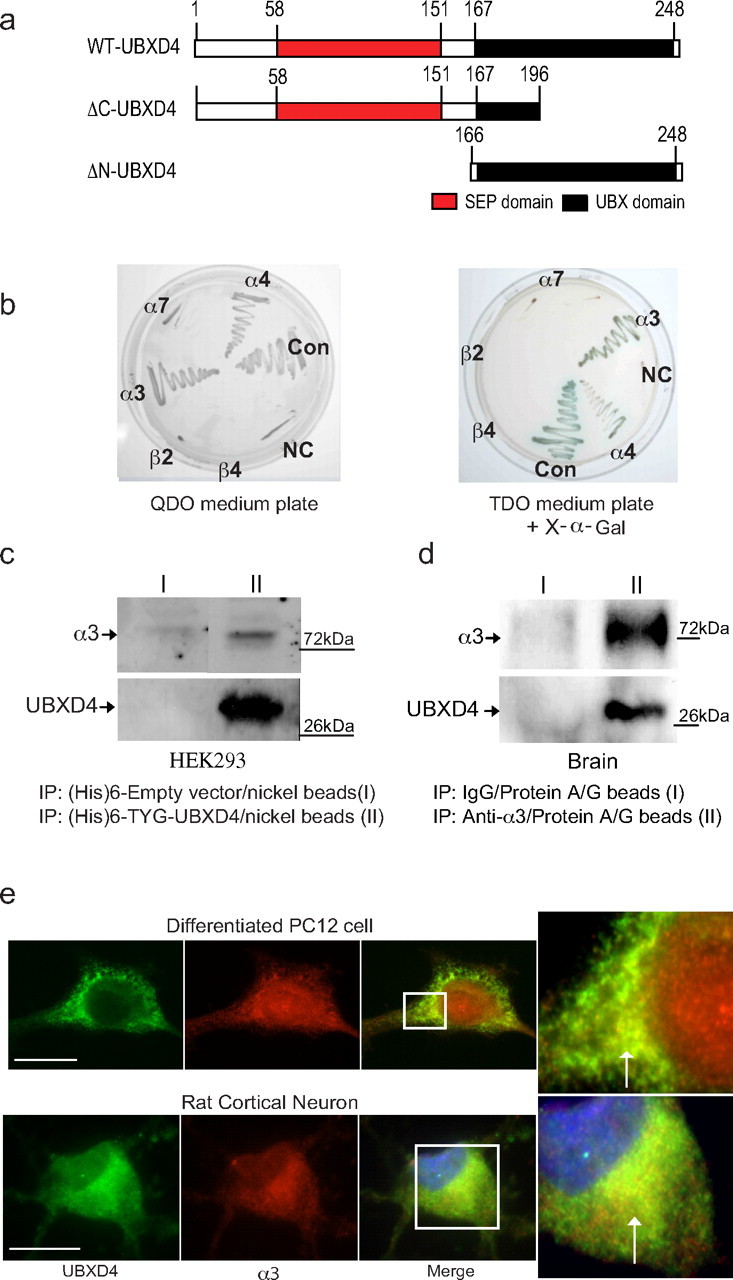
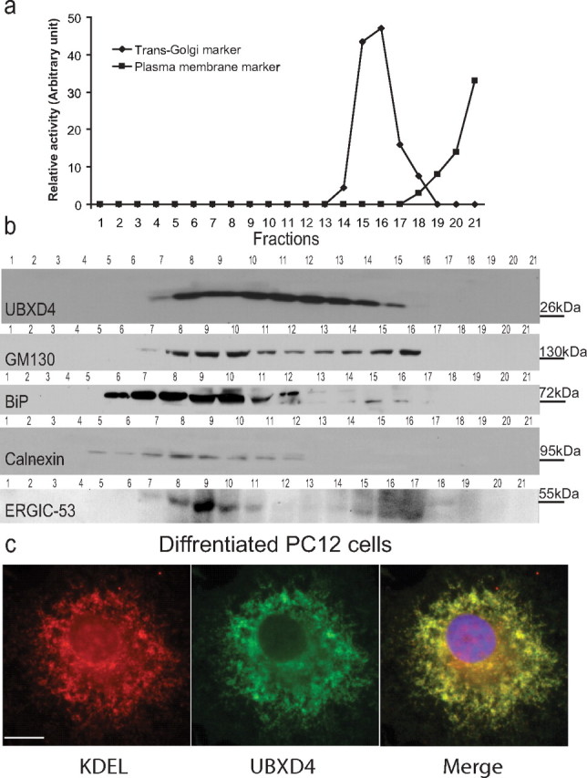
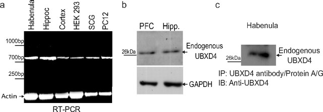
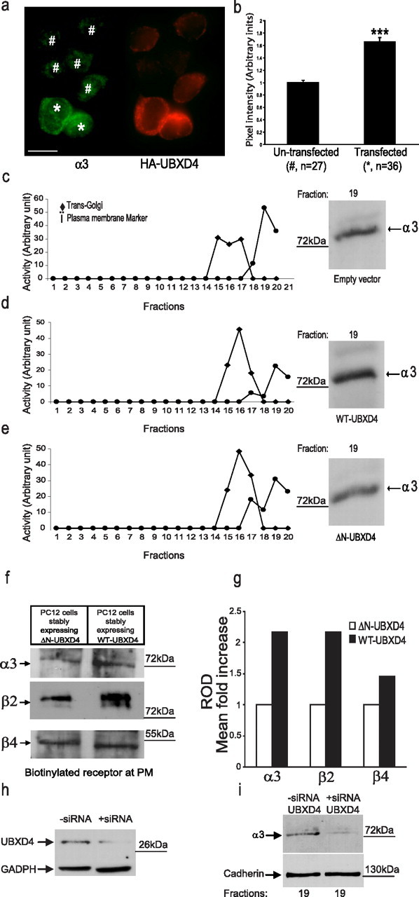
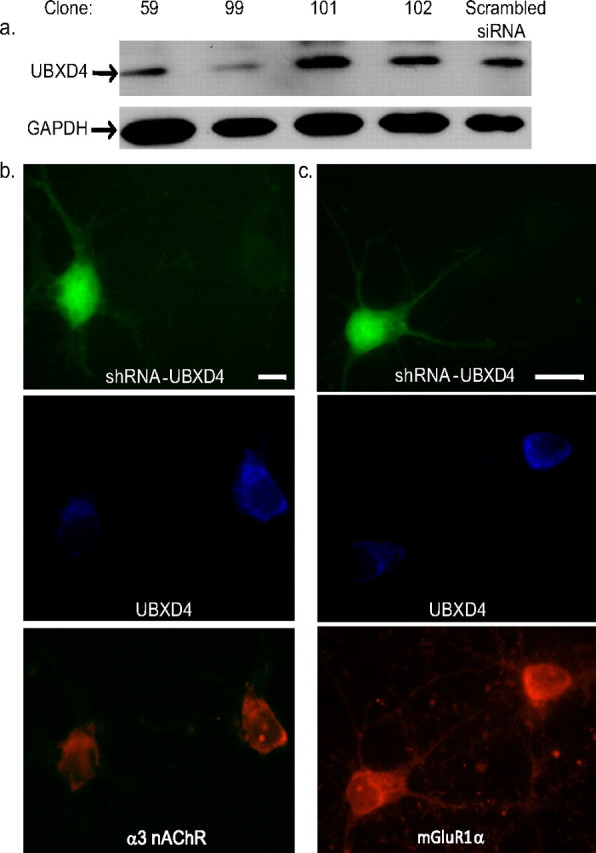
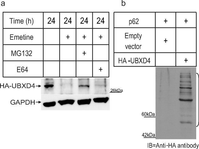
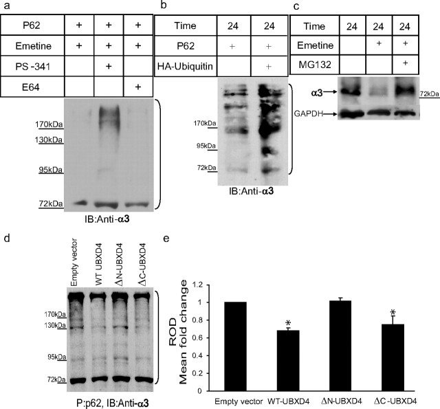
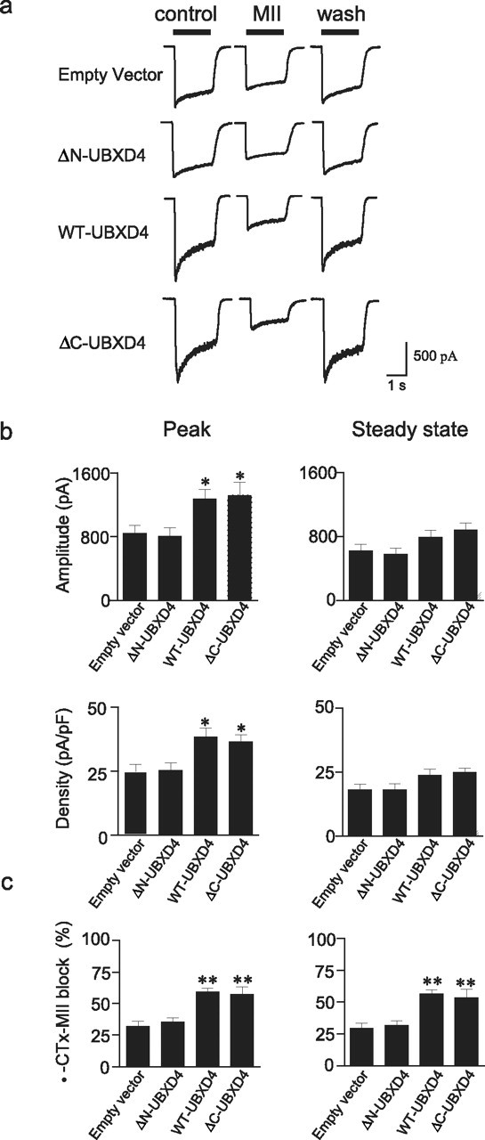
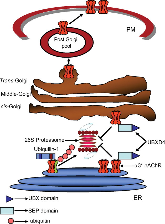
Adaptor proteins are likely to modulate spatially and temporally the trafficking of a number of membrane proteins, including neuronal nicotinic acetylcholine receptors (nAChRs). A yeast two-hybrid screen identified a novel UBX-containing protein, UBXD4, as one of the cytosolic proteins that interact directly with the alpha3 and alpha4 nAChR subunits. The function of UBX-containing proteins is largely unknown. Immunoprecipitation and confocal microscopy confirmed the interaction of UBXD4 with alpha3-containing nAChRs (alpha3* nAChRs) expressed in HEK293 cells, PC12 cells, and rat cortical neurons. Overexpression of UBXD4 in differentiated PC12 cells (dPC12) increased nAChR cell surface expression, especially that of the alpha3beta2 subtype. These findings were corroborated by electrophysiology, immunofluorescent staining, and biotinylation of surface receptors. Silencing of UBXD4 led to a significant reduction of alpha3* nAChRs in rat cortical neurons and dPC12 cells. Biochemical and immunofluorescence studies of endogenous UBXD4 showed that the protein is located in both the ER and cis-Golgi compartments. Our investigations also showed that the alpha3 subunit is ubiquitinated and that UBXD4 can interfere with its ubiquitination and consequent degradation by the proteasome. Our data suggest that UBXD4 modulates the distribution of alpha3* nAChRs between specialized intracellular compartments and the plasma membrane. This effect is achieved by controlling the stability of the alpha3 subunit and, consequently, the number of receptors at the cell surface.


Figures

Figure 1.
Figure 1.
UBXD4 interacts with α3* and α4* nAChRs. A yeast two-hybrid screen showed that the large intracellular loop of the α3 nAChR subunit interacts with a novel UBX-containing protein named UBXD4. a, Schematic diagrams illustrating the structural location of the UBX and SEP domains in WT-UBXD4 (top panel). Middle and bottom panels represent the original clone 42 found in Y2H (ΔC-UBXD4, middle) and the generated ΔN-UBXD4 clone lacking the SEP domain (bottom). b, Interaction was verified with cotransformation of α3 and UBXD4 into the Y187 yeast strain. Further experiments in yeast showed that UBXD4 interacts with α4 but not with the α7, β2, and β4 nAChR subunits demonstrating that UBXD4 is a selective partner for the α3 and α4 subunits. Left panel, Quadruple dropout media (−Leu/−His/−Trp/−Ade). Right panel, Triple dropout media (−Leu/−His/−Trp) containing X-α-gal for α-galactosidase activity; CON, positive control; NC, negative control. c, In vitro experiments with charged Ni+ columns showed that the α3 subunit can bind to (His)6-TYG-tagged UBXD4 protein expressed in HEK293 cells. d, UBXD4 interacts with native α3 nAChR subunits from mouse brain. PFC lysates were incubated either with rabbit IgG (I, control) or the anti-α3 antibody (II) immobilized on protein A/G. Eluted proteins were subjected to SDS-PAGE and immunoblotted with the anti-α3 and anti-UBXD4 antibodies. e, Differentiated PC12 cells endogenously expressing α3-containing nAChRs (top) or rat cortical neurons (bottom) were cultured on coverslips, and processed for immunocytochemistry. Staining of the permeabilized cells for UBXD4 (green) and α3 (red) demonstrated partial overlapping of UBXD4 and α3 in the perinuclear region of the cells, as indicated by the white arrows in the magnified inset. Nonspecific binding or staining of the nucleoplasm in PC12 cells was observed by omitting the primary antibody during the incubation process (Fujiwara et al., 2006). Confocal scale bar, 10 μm.
Figure 2.
Figure 2.
UBXD4 is located in the ER/Golgi compartments. a, b, Naive PC12 cells were subjected to 8–34% linear iodixanol gradient analysis. According to the leucine aminopeptidase (trans-Golgi marker) and alkaline phosphodiesterase (PM marker) activities and staining with anti-BiP and anti-calnexin (ER markers), anti-GM130 (cis- and middle Golgi marker), and ERGIC-53 (ERGIC marker) antibodies, UBXD4 is preferentially located in the ER/Golgi compartments (fractions 7–15). c, The predominant colocalization of UBXD4 with KDEL protein (an ER and cis-Golgi marker) in differentiated PC12 cells confirms that UBXD4 is an ER/Golgi protein. Confocal scale bar is 10 μm.
Figure 3.
Figure 3.
UBXD4 is expressed in neuronal and non-neuronal tissues. a, RT-PCR experiments showed that UBXD4 mRNA is expressed in all the examined tissues including cortex, hippocampus, habenula, peripheral ganglia, and two different cell lines (HEK293 and PC12 cells). b, c, Analysis of UBXD4 at the protein level with anti-UBXD4 antibodies demonstrated that the prefrontal cortex, hippocampus, and habenula express UBXD4 endogenously. Due to the small size of the habenula, we used IP techniques to enrich for UBXD4 by pulling down UBXD4 with anti-UBXD4 antibodies immobilized on protein A/G beads followed by probing with anti-UBXD4 (see Materials and Methods for details).
Figure 4.
Figure 4.
UBXD4 upregulates plasma membrane levels of α3* nAChRs. a, The fluorescent signal for α3 (green) increased when differentiated PC12 cells were transiently transfected with HA-tagged UBXD4 (red). The left panel shows the levels of α3 signal at the PM of UBXD4-overexpressing cells (*) compared with those of cells expressing endogenous levels of UBXD4 (#). Expression levels of UBXD4 where detected in the same cells with an anti-HA antibody after permeabilization (right panel). Confocal scale bar, 10 μm. b, Densitometric analysis of green pixel density in 63 cells (27 untransfected and 36 transfected) showed a significant increase in the total levels of α3 (p < 0.001). c–e, Differentiated PC12 cells stably expressing the corres-ponding empty vector (c), WT-UBXD4 (d), or ΔN-UBXD4 (e) were subjected to 8–34% linear iodixanol gradient fractionation. Enzymatic activity assays following iodixanol gradient fractionation were used to identify the fractions containing the Golgi and PM compartments (left panels). The fraction containing the highest levels of PM (fraction 19) showed increased levels of α3 upon overexpression of UBXD4 (right panels). ΔN-UBXD4 overexpression had no effect on α3 PM levels. f, Biotinylation experiments confirmed that expression of WT-UBXD4 but not ΔN-UBXD4 (control) greatly increases the surface levels of the α3 and β2 nAChR subunits. g, Quantitation of the data shown in f indicated that α3 and β2 levels increased 2.2-fold while β4 levels increased 1.4-fold compared with control. h, Stable expression of UBXD4 siRNA in PC12 cells decreased UBXD4 expression. i, The reduction in UBXD4 levels led to a significant decrease in α3 levels in PM fractions. The above experiments were conducted twice with similar results each time.
Figure 5.
Figure 5.
UBXD4 shRNA selectively decreases α3 nAChRs signal in rat cortical neurons. a, Western blot analyses of PC12 cells transfected with four lentiviral-based shRNA showed that clone 99 can efficiently silence endogenous UBXD4. b, Confocal microscopy of rat cortical neurons transfected with clone 99 against UBXD4 demonstrated a significant reduction of α3 signal in permeabilized, shRNA-transfected cells. The reduction of signal for α3-containing nAChRs by UBXD4 siRNA suggests that UBXD4 regulates trafficking of α3. c, shRNA against UBXD4 had no significant effect on mGluR1α receptor levels detected in rat cortical neuron. See supplemental Figure 5, c and d (available at www.jneurosci.org as supplemental material), for the quantification of the confocal data.
Figure 6.
Figure 6.
UBXD4 is ubiquitinated and degraded by the 26S proteasome. a, A 24 h exposure to the proteasome blocker MG132 in the presence of the protein synthesis blocker, emetine, decreased the rate of degradation of UBXD4 in HEK293 cells transiently transfected with HA-tagged UBXD4. Exposure to the lysosome inhibitor E64 did not alter the degradation rate of UBXD4. b, A ladder of ubiquitinated, HA-tagged UBXD4 could be pulled down with p62 from α3β2 expressing HEK293 cells transiently transfected with HA-tagged UBXD4, but not with the HA-empty vector. The bracket on the right marks a ladder of bands corresponding to polyubiquitinated UBXD4. The above experiments were conducted twice with similar results each time.
Figure 7.
Figure 7.
UBXD4 decreases the levels of ubiquitinated α3. Differentiated PC12 cells were treated for 24 h with PS-341 (a selective proteasome inhibitor) or E64 (an inhibitor of the lysosome) in the presence of emetine (a protein synthesis blocker). Cell lysates were probed for α3. a, A ladder of various polyubiquitinated forms of α3 (bracket) could be pulled down by the p62-derived UBA domain, suggesting that α3 is ubiquitinated and can be degraded by the proteasome. In untreated cells (left lane) and cells treated with E64 (right lane), the majority of α3 receptors was degraded by the proteasome after ubiquitination. b, In another set of experiments, the level of ubiquitination of α3 in the presence of overexpressed HA-ubiquitin was determined. Cell lysates of PC12 cells stably expressing empty vector or HA-ubiquitin were subjected to p62 pull down followed by immunoblotting with anti-α3 antibodies. Overexpression of HA-ubiquitin led to an increase in the levels of ubiquitinated α3. c, The degradation of the α3 nAChR subunit was decreased dramatically in the presence of the proteasome inhibitor MG132 in PC12 cells. d, P62 pull down was performed on cell lysates from dPC12 cells stably transfected with the corresponding empty vector, WT-UBXD4, ΔN-UBXD4, or ΔC-UBXD4 followed by Western blot analysis with anti-α3 antibodies. A ladder of ubiquitinated α3 could be detected in each sample. Quantification of the α3 signal showed that WT-UBXD4 and ΔC-UBXD4, but not ΔN-UBXD4, significantly decrease the ubiquitinated levels of α3 (e; p < 0.05). The brackets on the right (a, b, d) mark a ladder of bands corresponding to polyubiquitinated α3.
Figure 8.
Figure 8.
Functional regulation of α3* nAChRs by UBXD4. a, Currents elicited by 300 μm ACh were measured before, during, and after a 5 min exposure to 100 nm α-CTx-MII in dPC12 cells stably transfected with empty vector, ΔN-UBXD4, WT-UBXD4, or ΔC-UBXD4. b shows peak (left panels) and steady-state (right panels) current amplitude and density in the various cell lines described in a. Overexpression of WT-UBXD4 and ΔC-UBXD4 produced a significant increase (p < 0.05) in peak amplitude and current density compared with empty vector or ΔN-UBXD4. c, Block by α-CTx-MII of peak (left panel) and steady-state (right panel) α3* nAChR currents. The α-CTx-MII-sensitive component was greater in cells overexpressing WT-UBXD4 and ΔC-UBXD4 than in cells expressing the corresponding empty vector or ΔN-UBXD4 (p < 0.01). Values represent mean ± SEM values from cells expressing empty vector (n = 16), ΔN-UBXD4 (n = 12), WT-UBXD4 (n = 34), and/or ΔC-UBXD4 (n = 8), respectively.
Figure 9.
Figure 9.
UBXD4 facilitates the trafficking of α3* nAChRs. Model showing the putative role of UBXD4 in the trafficking of α3* nAChRs after assembly in the endoplasmic reticulum (ER). Unfolded subunits and unmasked receptors are recognized by the ER-associated degradation system (ERAD) with the aid of chaperons like Plic-1. Association of other adaptor proteins such as UBXD4 can mask the degradation signal allowing nAChRs to bypass the checkpoints at the ER and cis-Golgi level before insertion in the PM.

Similar articles

Cited by

References

    1. Alexandru G, Graumann J, Smith GT, Kolawa NJ, Fang R, Deshaies RJ. UBXD7 binds multiple ubiquitin ligases and implicates p97 in HIF1alpha turnover. Cell. 2008;134:804–816. - PMC - PubMed
    1. Arias HR, Bhumireddy P. Anesthetics as chemical tools to study the structure and function of nicotinic acetylcholine receptors. Curr Protein Pept Sci. 2005;6:451–472. - PubMed
    1. Arikkath J, Israely I, Tao Y, Mei L, Liu X, Reichardt LF. Erbin controls dendritic morphogenesis by regulating localization of δ-catenin. J Neurosci. 2008;28:7047–7056. - PMC - PubMed
    1. Bedford FK, Kittler JT, Muller E, Thomas P, Uren JM, Merlo D, Wisden W, Triller A, Smart TG, Moss SJ. GABA(A) receptor cell surface number and subunit stability are regulated by the ubiquitin-like protein Plic-1. Nat Neurosci. 2001;4:908–916. - PubMed
    1. Belizario JE, Alves J, Garay-Malpartida M, Occhiucci JM. Coupling caspase cleavage and proteasomal degradation of proteins carrying PEST motif. Curr Protein Pept Sci. 2008;9:210–220. - PubMed

Publication types

MeSH terms