. 2009 May 27;29(21):7065-78.
doi: 10.1523/JNEUROSCI.6072-08.2009.

Realignment of interaural cortical maps in asymmetric hearing loss

Affiliations

Realignment of interaural cortical maps in asymmetric hearing loss

Steven W Cheung et al. J Neurosci. .

Abstract

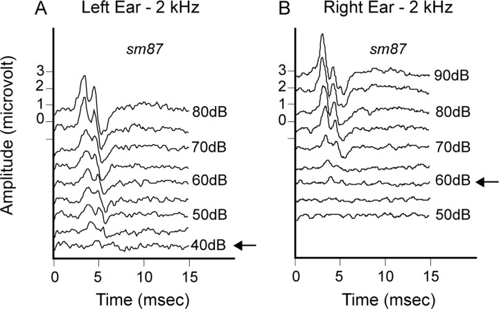
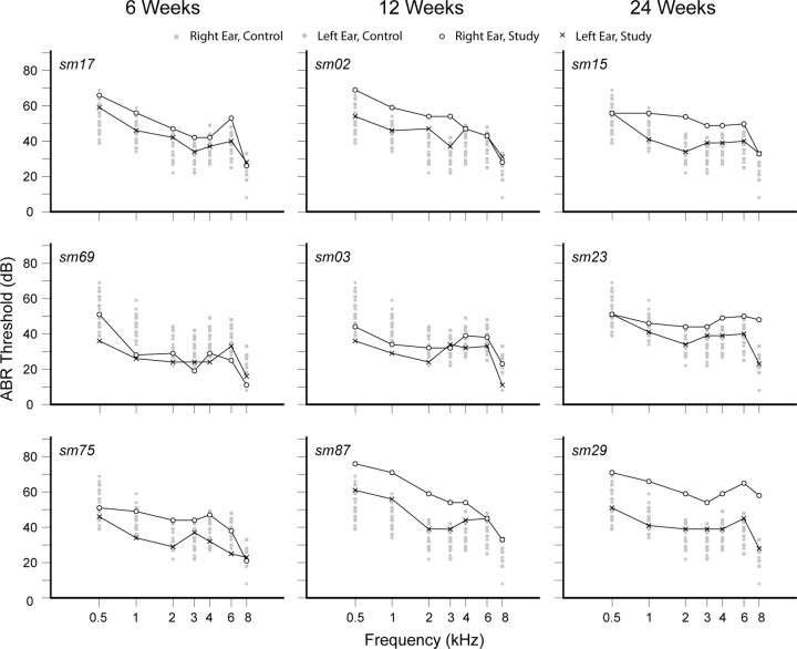
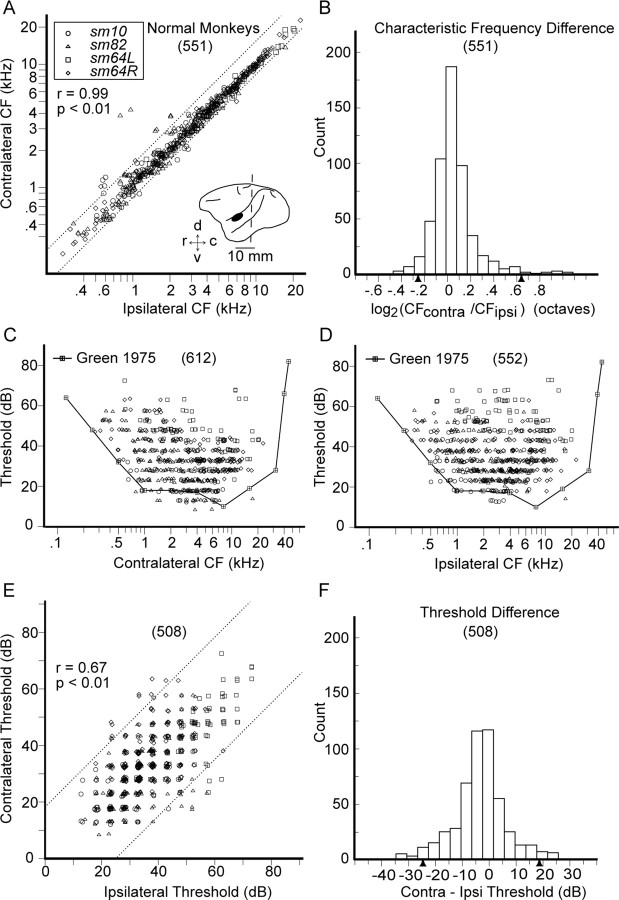
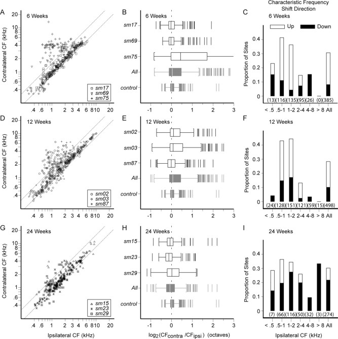
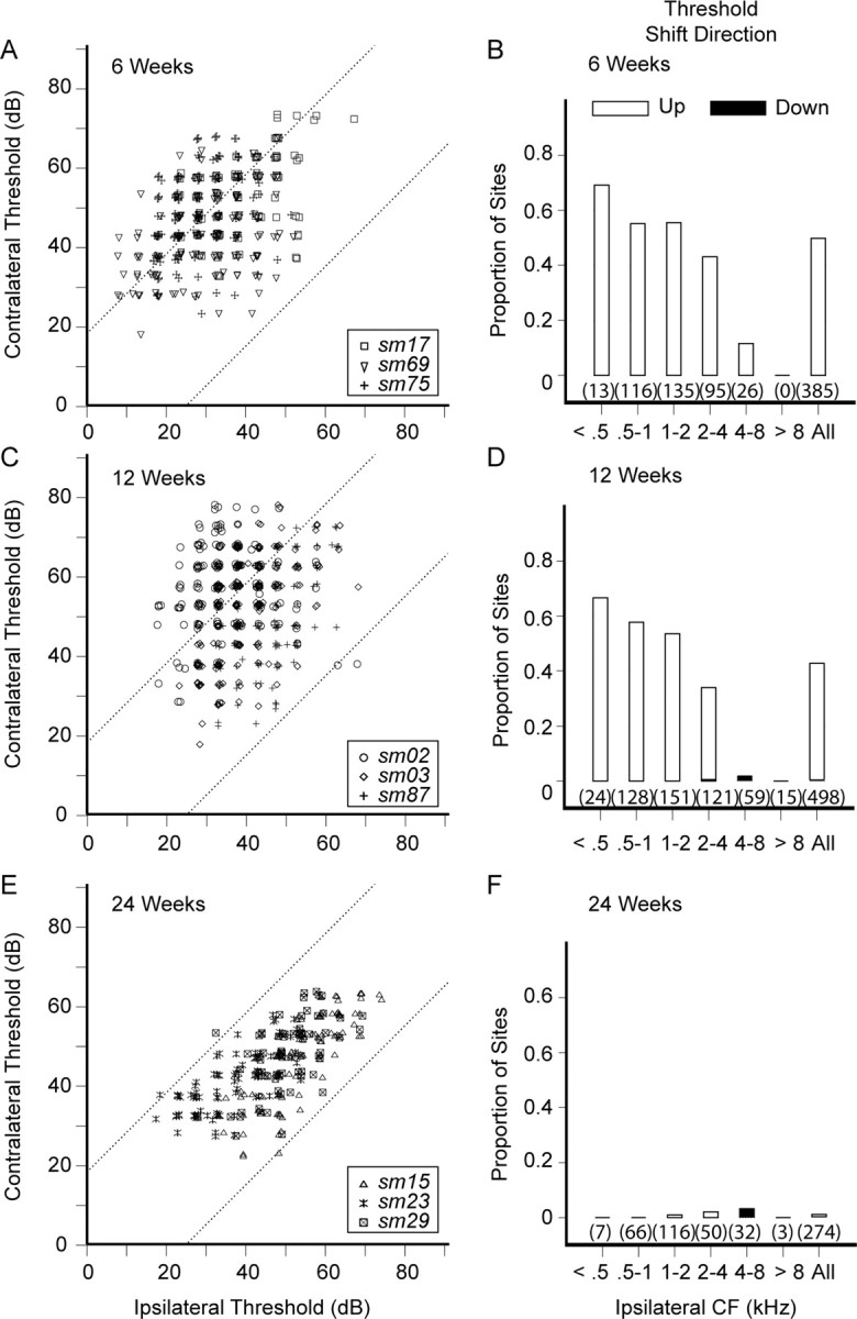
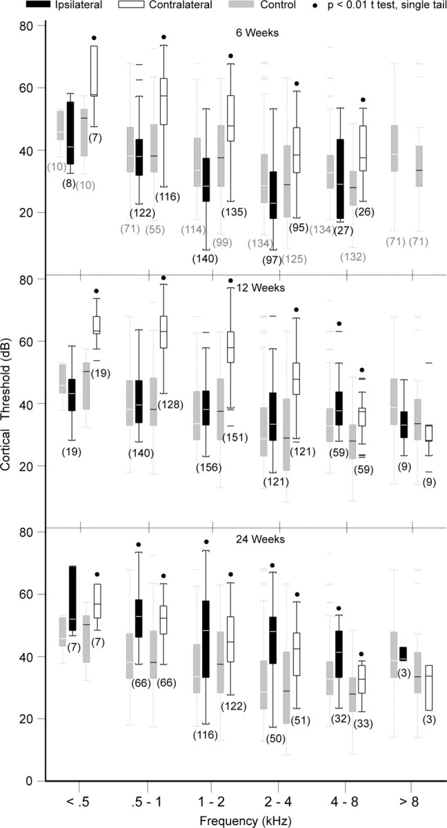
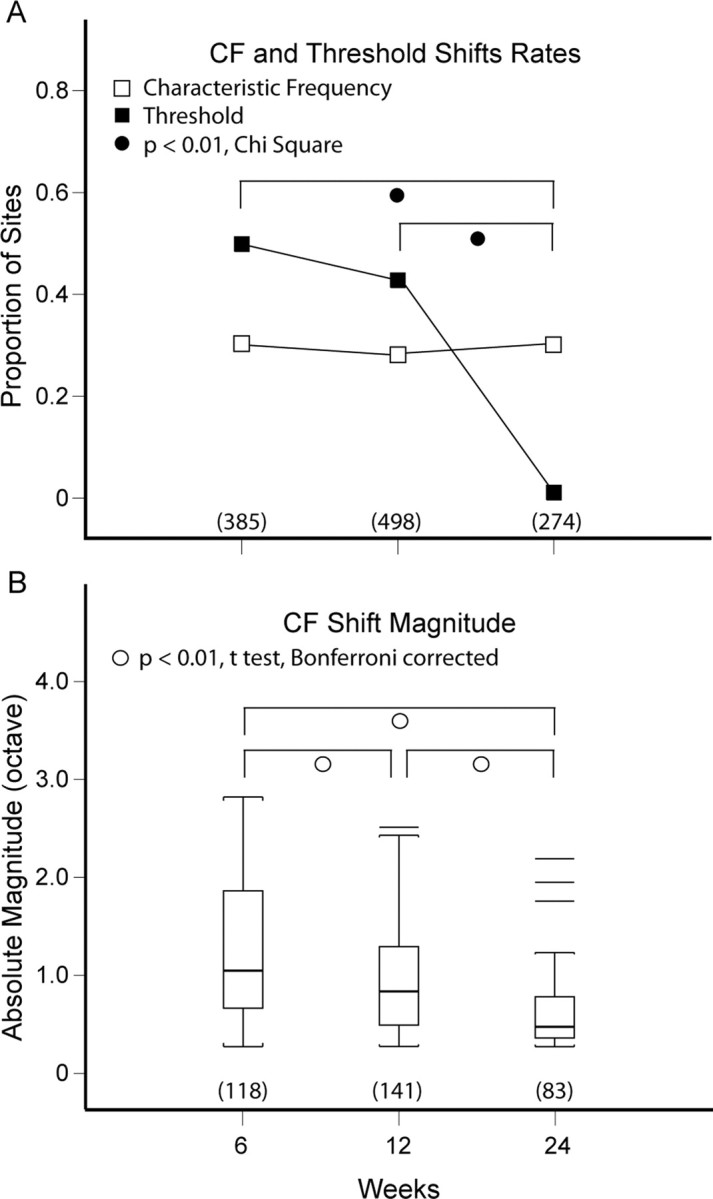
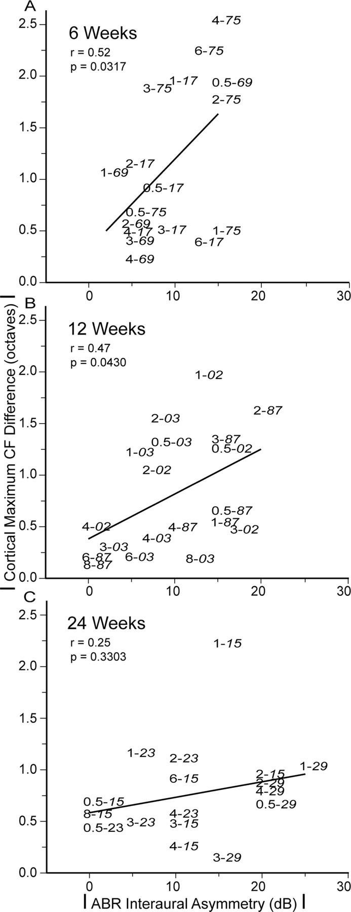
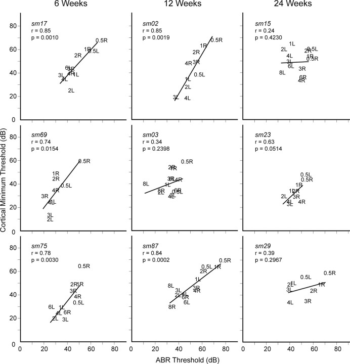
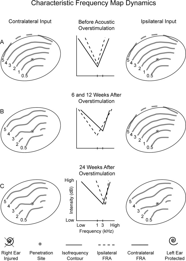
Misalignment of interaural cortical response maps in asymmetric hearing loss evolves from initial gross divergence to near convergence over a 6 month recovery period. The evolution of left primary auditory cortex (AI) interaural frequency map changes is chronicled in squirrel monkeys with asymmetric hearing loss induced by overstimulating the right ear with a 1 kHz tone at 136 dB for 3 h. AI frequency response areas (FRAs), derived from tone bursts presented to the poorer or better hearing ears, are compared at 6, 12, and 24 weeks after acoustic overstimulation. Characteristic frequency (CF) and minimum threshold parameters are extracted from FRAs, and they are used to quantify interaural response map differences. A large interaural CF map misalignment of DeltaCF approximately 1.27 octaves at 6 weeks after overstimulation decreases substantially to DeltaCF approximately 0.62 octave at 24 weeks. Interaural cortical threshold map misalignment faithfully reflects peripheral asymmetric hearing loss at 6 and 12 weeks. However, AI threshold map misalignment essentially disappears at 24 weeks, primarily because ipsilateral cortical thresholds have become unexpectedly elevated relative to peripheral thresholds. The findings document that plastic change in central processing of sound stimuli arriving from the nominally better hearing ear may account for progressive realignment of both interaural frequency and threshold maps.


Figures

Figure 1.
Figure 1.
ABR waveforms for threshold determination at 2 kHz. A, Left ear. B, Right ear. Arrows indicate threshold.
Figure 2.
Figure 2.
ABR audiograms for study monkeys after acoustic overstimulation with a 1 kHz tone at 136 dB for 3 h directed at the right ear. Overall mild asymmetric sensorineural hearing loss. Gray symbols (square, right ear; diamond, left ear) are thresholds for normal monkeys.
Figure 3.
Figure 3.
Characteristic frequency and threshold data for contralateral versus ipsilateral ear input from four cortices in three normal squirrel monkeys. A, Relationship between ipsilateral and contralateral CFs. R, Right AI; L, left AI. Inset, Squirrel monkey brain caricature. The general recording area for AI on the temporal gyrus is ventral to the lateral sulcus and 4–8 mm rostral to the interaural line (dotted). AI varies in its rostral-caudal location and size on the temporal gyrus. r, Rostral; c, caudal; v, ventral; d, dorsal. B, CF difference distribution in octaves. C, D, Population contralateral and ipsilateral neuronal thresholds superimposed on behavioral thresholds by Green (1975). The lowest neuronal thresholds are in close proximity to behavioral thresholds. For CF >8 kHz, neuronal sampling is more sparse. E, Relationship between ipsilateral and contralateral thresholds. F, Threshold difference distribution in decibels. Solid triangles mark upper and lower tail limits for α/2 = 0.025 for both empirical distributions. r = correlation coefficient. Sample size is in parentheses.
Figure 4.
Figure 4.
Characteristic frequency for contralateral versus ipsilateral ear input in squirrel monkeys with mild asymmetric hearing loss. A, D, G, The 6, 12, and 24 week recovery time periods. Interaural CF misalignment is evident. There is a reversal of shift direction from mostly higher values (CF shift up) at 6 and 12 weeks to lower values (CF shift down) at 24 weeks. Dotted lines indicate boundaries of the 95% confidence interval derived from empirical difference distributions. B, E, H, CF misalignment variations among monkeys in each study group. Tukey box plots show extended upper and/or lower tails in study monkeys that account for CF shift up and CF shift down neurons. At 6 weeks, monkey sm75 is the largest contributor to the group's extended tails. At 12 weeks, all three monkeys contribute evenly to both extended tails. At 24 weeks, all three monkeys also contribute evenly to both extended tails, but the CF shift down tail predominates. C, F, I, Proportion of sites showing either CF shift up or shift down for each study group. Data have been segmented to single octave bands referenced to the ipsilateral CF. The total proportion of sites showing interaural CF difference is ∼30% for all three recovery time periods, but the dominant direction of CF shift reverses at 24 weeks. Sample size is in parentheses.
Figure 5.
Figure 5.
Threshold for contralateral versus ipsilateral ear input in squirrel monkeys with mild asymmetric hearing loss. A, C, E, The 6, 12, and 24 week recovery time periods. Threshold shift direction is almost exclusively to higher values (threshold shift up) at 6 and 12 weeks, an expected result from asymmetric injury to the auditory periphery. Threshold shift becomes inapparent by 24 weeks, an unexpected finding. B, D, F, Corresponding recovery time periods for the proportion of sites showing either threshold shift up or shift down. Data have been segmented to single octave bands referenced to the ipsilateral CF. The total proportion of sites showing interaural threshold difference is ∼45% for 6 and 12 weeks, but drops to <1% by 24 weeks. Sample size is in parentheses.
Figure 6.
Figure 6.
Cortical threshold Tukey box plots for squirrel monkeys with mild asymmetric hearing loss segmented by single octave ipsilateral CF bands. 6 Weeks and 12 Weeks, Contralateral thresholds are elevated, whereas ipsilateral thresholds are indistinguishable from controls for ipsilateral CF <4 kHz. 24 Weeks, Both contralateral and ipsilateral thresholds are elevated.
Figure 7.
Figure 7.
Summary of CF and threshold shift rates, and CF shift magnitude. A, CF shift rate, up or down, is ∼30% for all three recovery time periods. Threshold shift rates are indistinguishable (∼45%) at 6 and 12 weeks, but they differ from the threshold shift rate at 24 weeks (<1%). B, CF shift magnitude declines significantly (p < 0.01) with each successive recovery time period.
Figure 8.
Figure 8.
Relationship between absolute ABR interaural threshold asymmetry and maximum CF difference levels for all recovery time periods. A, B, Monkeys at 6 and 12 weeks show moderately strong linear correlation (r = 0.52, 6 weeks; r = 0.47, 12 weeks; both p < 0.05). C, Monkeys at 24 weeks do not show a significant linear relationship (p > 0.05).
Figure 9.
Figure 9.
Relationship between minimum cortical and corresponding ABR thresholds for both ears in individual study monkeys. 6 Weeks and 12 Weeks, Linear regression fits (p < 0.05) show strong correlation (r > 0.74) for five of six study monkeys. 24 Weeks, Linear regression fits are not significant (p > 0.05) for all three study monkeys.
Figure 10.
Figure 10.
Summary model description of frequency map dynamics in asymmetric mild sensorineural hearing loss. Left and right columns depict ipsilateral and contralateral isofrequency contour reorganization, and the middle column depicts FRA dynamics for CF and threshold. A, The first row shows that contralateral and ipsilateral CF maps are in alignment in normal monkeys. B, At 6 and 12 weeks after acoustic overstimulation, contralateral CF and threshold shifts are to mostly higher values. Ipsilateral FRA parameters remain similar to those in normal monkeys. Misalignment of interaural CF and threshold maps is evident. C, At 24 weeks, ipsilateral CF and threshold shifts to higher values emerge. Realignment of interaural CF and threshold maps follows. Dashed line (5 kHz isofrequency contour) indicates entry into the suprameatal plane.

References

    1. Barsz K, Wilson WW, Walton JP. Reorganization of receptive fields following hearing loss in inferior colliculus neurons. Neuroscience. 2007;147:532–545. - PMC - PubMed
    1. Benson DA, Teas DC. Single unit study of binaural interaction in the auditory cortex of the chinchilla. Brain Res. 1976;103:313–338. - PubMed
    1. Brainard MS, Knudsen EI. Experience-dependent plasticity in the inferior colliculus: a site for visual calibration of the neural representation of auditory space in the barn owl. J Neurosci. 1993;13:4589–4608. - PMC - PubMed
    1. Brainard MS, Knudsen EI. Dynamics of visually guided auditory plasticity in the optic tectum of the barn owl. J Neurophysiol. 1995;73:595–614. - PubMed
    1. Calford MB, Wang C, Taglianetti V, Waleszczyk WJ, Burke W, Dreher B. Plasticity in adult cat visual cortex (area 17) following circumscribed monocular lesions of all retinal layers. J Physiol. 2000;524:587–602. - PMC - PubMed

MeSH terms