. 2009 May 27;29(21):7092-7.
doi: 10.1523/JNEUROSCI.6062-08.2009.

Pannexin 1: the molecular substrate of astrocyte "hemichannels"

Affiliations

Pannexin 1: the molecular substrate of astrocyte "hemichannels"

Rodolfo Iglesias et al. J Neurosci. .

Abstract

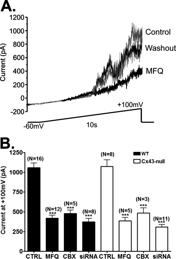
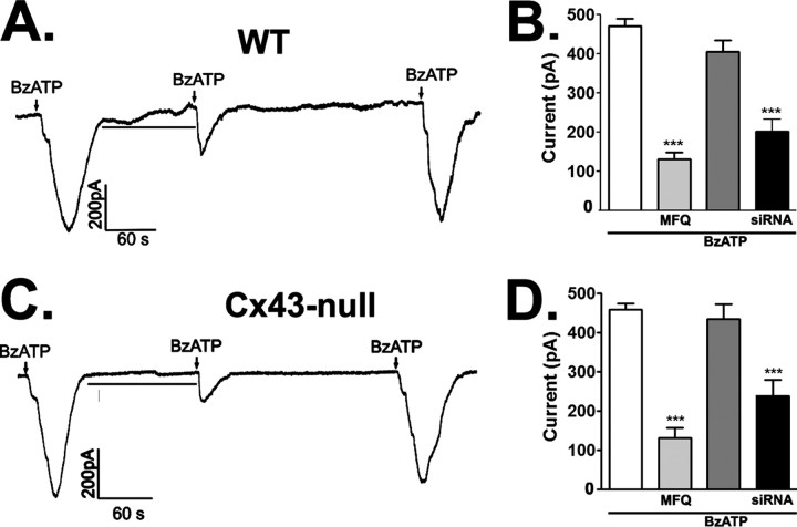
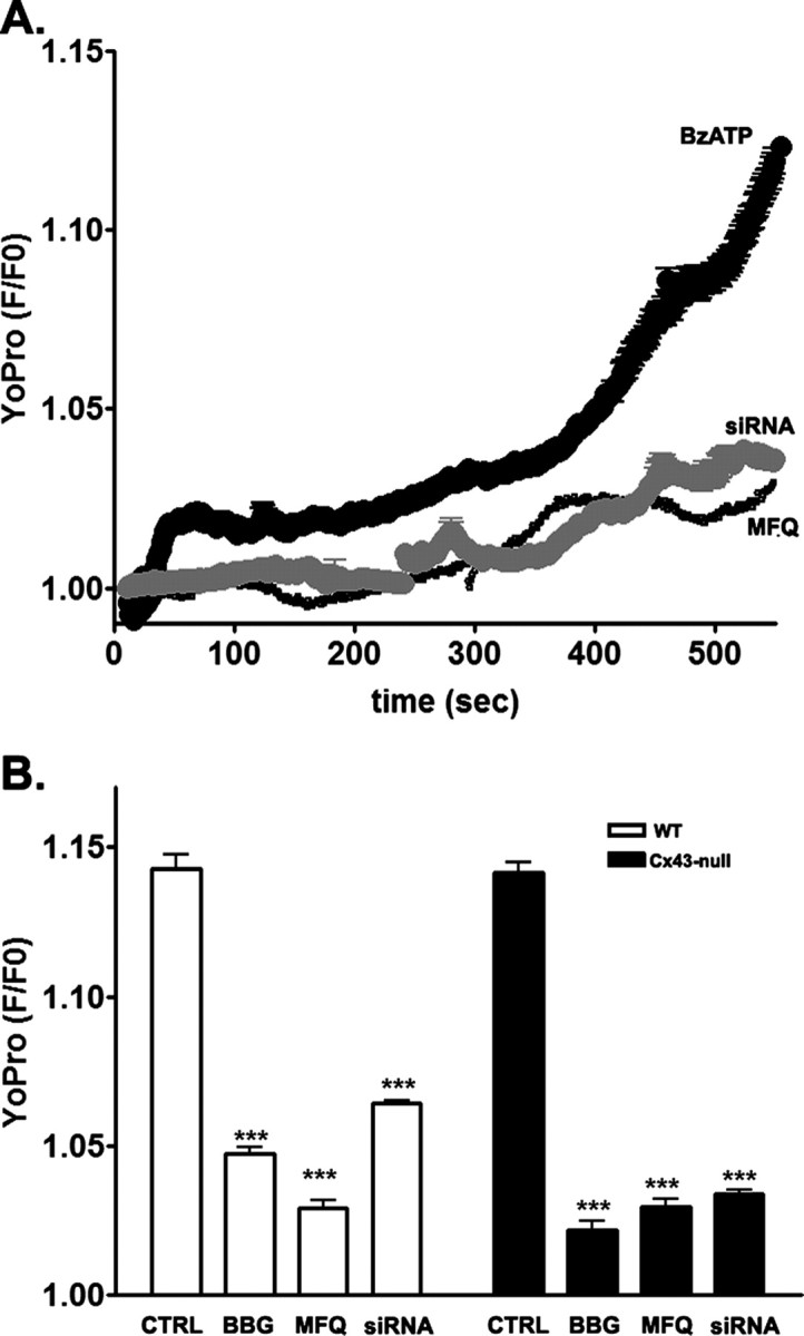
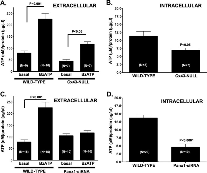
Purinergic signaling plays distinct and important roles in the CNS, including the transmission of calcium signals between astrocytes. Gap junction hemichannels are among the mechanisms proposed by which astrocytes might release ATP; however, whether the gap junction protein connexin43 (Cx43) forms these "hemichannels" remains controversial. Recently, a new group of proteins, the pannexins, have been shown to form nonselective, high-conductance plasmalemmal channels permeable to ATP, thereby offering an alternative for the hemichannel protein. Here, we provide strong evidence that, in cultured astrocytes, pannexin1 (Panx1) but not Cx43 forms hemichannels. Electrophysiological and fluorescence microscope recordings performed in wild-type and Cx43-null astrocytes did not reveal any differences in hemichannel activity, which was mostly eliminated by treating Cx43-null astrocytes with Panx1-short interfering RNA [Panx1-knockdown (Panx1-KD)]. Moreover, quantification of the amount of ATP released from wild-type, Cx43-null, and Panx1-KD astrocytes indicates that downregulation of Panx1, but not of Cx43, prevented ATP release from these cells.


Figures

Figure 1.
Figure 1.
Effects of gap junction channel blockers on voltage-activated Panx1 currents in astrocytes. A, Examples of outward currents recorded from a single astrocyte in the absence and presence of MFQ (100 nm) and after MFQ washout. Currents were obtained in response to voltage ramps (−60 to +100 mV) with −60 mV holding membrane potential. B, Bar histograms showing the mean ± SE values of current amplitudes at the end of the current ramps (+100 mV) recorded from WT (black bars) and Cx43-null (white bars) astrocytes bathed in control (CTRL) solution and solutions containing CBX (50 μm) and MFQ (100 nm). Current amplitudes obtained for astrocytes treated for 48 h with Panx1 siRNA are shown in the last bars. The numbers in parentheses correspond to the number of cells tested. ***p < 0.01.
Figure 2.
Figure 2.
P2X7R-induced Panx1 currents in astrocytes. A, C, Representative current traces induced by 5 s application of BzATP (50 μm) in WT (A) and Cx43-null (C) astrocytes are shown to be greatly reduced by 5 min preincubation (lines) with 100 nm MFQ; after washout of MFQ, agonist induced currents of similar amplitudes as those recorded after the first application. B, D, Bar histograms show the mean ± SE values (N = 4–10 cells) corresponding to data displayed in A and C, respectively. The traces were obtained by holding the membrane at −60 mV. ***p < 0.01.
Figure 3.
Figure 3.
The P2X7R–pannexin 1 complex mediates astrocyte membrane permeabilization. A, Representative time course of YoPro uptake recorded from WT astrocytes exposed to BzATP (300 μm) in the absence and presence of MFQ (100 nm) and after Panx1 knockdown with siRNA. B, Bar histograms showing the mean ± SE values of the relative YoPro fluorescence intensity obtained for wild-type (white bars) and Cx43-null (black bars) astrocytes treated for 500 s with BzATP (300 μm) in the absence and presence of 1 μm BBG and of 100 nm MFQ. ***p < 0.01.
Figure 4.
Figure 4.
BzATP-induced ATP release from astrocytes. Bar histograms showing the mean ± SE values of ATP (A, C) present in the extracellular solution in response to BzATP prestimulation of WT and Cx43-null (A) and WT and Panx1-siRNA-treated (C) astrocytes. B and D show the mean ± SE values of intracellular ATP present in the cytosol of WT and Cx43-null astrocytes (B) and in cells untreated and treated with Panx1-siRNA (C). Note that, in A and C, the values of ATP are expressed as nanomolar, and in B and D, in micromolar.

References

    1. Bao L, Locovei S, Dahl G. Pannexin membrane channels are mechanosensitive conduits for ATP. FEBS Lett. 2004;572:65–68. - PubMed
    1. Bruzzone R, Hormuzdi SG, Barbe MT, Herb A, Monyer H. Pannexins, a family of gap junction proteins expressed in brain. Proc Natl Acad Sci U S A. 2003;100:13644–13649. - PMC - PubMed
    1. Bruzzone R, Barbe MT, Jakob NJ, Monyer H. Pharmacological properties of homomeric and heteromeric pannexin hemichannels expressed in Xenopus oocytes. J Neurochem. 2005;92:1033–1043. - PubMed
    1. Bukauskas FF, Bukauskiene A, Verselis VK. Conductance and permeability of the residual state of connexin43 gap junction channels. J Gen Physiol. 2002;119:171–185. - PMC - PubMed
    1. Contreras JE, Sánchez HA, Eugenin EA, Speidel D, Theis M, Willecke K, Bukauskas FF, Bennett MV, Sáez JC. Metabolic inhibition induces opening of unapposed connexin 43 gap junction hemichannels and reduces gap junctional communication in cortical astrocytes in culture. Proc Natl Acad Sci U S A. 2002;99:495–500. - PMC - PubMed

Publication types

MeSH terms

LinkOut - more resources