. 2009 Jul;37(13):4498-507.
doi: 10.1093/nar/gkp437. Epub 2009 May 27.

High-affinity triplex targeting of double stranded DNA using chemically modified peptide nucleic acid oligomers

Affiliations

High-affinity triplex targeting of double stranded DNA using chemically modified peptide nucleic acid oligomers

Mads E Hansen et al. Nucleic Acids Res. 2009 Jul.

Abstract

While sequence-selective dsDNA targeting by triplex forming oligonucleotides has been studied extensively, only very little is known about the properties of PNA-dsDNA triplexes--mainly due to the competing invasion process. Here we show that when appropriately modified using pseudoisocytosine substitution, in combination with (oligo)lysine or 9-aminoacridine conjugation, homopyrimidine PNA oligomers bind complementary dsDNA targets via triplex formation with (sub)nanomolar affinities (at pH 7.2, 150 mM Na(+)). Binding affinity can be modulated more than 1000-fold by changes in pH, PNA oligomer length, PNA net charge and/or by substitution of pseudoisocytosine for cytosine, and conjugation of the DNA intercalator 9-aminoacridine. Furthermore, 9-aminoacridine conjugation also strongly enhanced triplex invasion. Specificity for the fully matched target versus one containing single centrally located mismatches was more than 150-fold. Together the data support the use of homopyrimidine PNAs as efficient and sequence selective tools in triplex targeting strategies under physiological relevant conditions.


Figures

Figure 1.
Figure 1.
Comparison of PNA oligomer and TFO binding to the complementary dsDNA target at pH 6.3. (A) Autoradiograph showing DNase I footprint formed by PNA1846 (lanes 3–9) and TFO1 (lanes 11–17) bound to the complementary dsDNA target from p322. The position of the sequence target is indicated adjacent to the A/G sequence reaction (lane 1). Asterisk indicates the position of the 32P-label. Lanes 2 and 10 are controls without oligomer. (B) Quantitative analysis of the data shown in (A). Percentage DNaseI protection as a function of PNA1846 or TFO1 concentration. (C) Autoradiograph showing PNA1846 binding to the complementary sequence target in p322 as analysed by gel-shift analysis. The following PNA1846 concentrations (µM) were used: w/o (lane 1), 0.002 (lane 2), 0.006 (lane 3), 0.02 (lane 4), 0.06 (lane 5), 0.17 (lane 6), 0.5 (lane 7), 1.5 (lane 8) and 4.5 (lane 9). (D) Percentage PD-complex (triplex or invasion) formed as a function of PNA concentration is shown. Free dsDNA is included for reference (indicated as duplex). Error bars indicate standard error of the mean (SEM) of six experimental repetitions. Notice the bell-shaped curve for triplex formation. Also notice the logarithmic scale of the x-axis.
Figure 2.
Figure 2.
Effect of pH on pentadecamer PNA-dsDNA triplex formation. The graph shows percentage PD triplex formed at the indicated pH as a function of the PNA1846 concentration upon binding to the complementary dsDNA sequence target as determined by gel-shift analysis. Error bars indicate SEM using three to four experimental repetitions, except for the data from pH 6.3 where the data from Figure 1C is included for reference. See Figure S3 for example of gel-shift data.
Figure 3.
Figure 3.
Effect of PNA length on PNA-dsDNA triplex formation at pH 6.3. The graph shows percentage PD triplex formed as a function of the PNA concentration upon binding to the complementary dsDNA target as determined by gel-shift analysis and using the indicated (+2) PNA oligomers. Error bars indicate SEM using two to three experimental repetitions, except for the data from pH 6.3 where the data from Figure 1C is included for reference. See Figure S4 for example of gel-shift data.
Figure 4.
Figure 4.
Effect of PNA net-charge on pentadecamer PNA–dsDNA triplex formation at pH 6.3. The graph shows percentage PD triplex formed as a function of the concentration of the (+1 to +5) PNA oligomers upon binding to the complementary dsDNA sequence as determined by gel-shift analysis using the indicated PNA oligomers. Error bars indicate SEM using two experimental repetitions, except for the data from pH 6.3 where the data from Figure 1C is included for reference. See Figures S5 and S3 for example of gel-shift data.
Figure 5.
Figure 5.
Triplex formation by J-base versus cytosine containing pentadecamer PNAs at pH 7.2. The graph shows percentage PD triplex formed as a function of the PNA concentration on binding to the complementary dsDNA target using the indicated PNA oligomers. The symbol nomenclature is similar to that given in Table 2, column 3. Error bars indicate SEM using two to three experimental repetitions. See Figures S6 and S3 for example of gel-shift data.
Figure 6.
Figure 6.
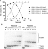
Triplex formation and triplex invasion by 9-aminoacridine modified pentadecamer PNAs at pH 7.2. (A) Graph showing percentage PD complex (triplex or invasion) formed as a function of the PNA concentration on binding to the complementary sequence target in p322. (B) Autoradiograph showing an example of gel-shift analysis of PNA3003 binding to dsDNA. The following PNA oligomer concentrations (µM) were used: w/o (lane 1), 0.002 (lane 2), 0.006 (lane 3), 0.02 (lane 4), 0.06 (lane 5), 0.17 (lane 6), 0.5 (lane 7), 1.5 (lane 8) and 4.5 (lane 9). The symbol nomenclature is similar to that given in Table 2, column 3. Error bars indicate SEM using two to three experimental repetitions.
Figure 7.
Figure 7.
Triplex formation and triplex invasion by 9-aminoacridine modified J-base pentadecamer PNAs at pH 7.2. (A) The graph shows percentage PD complex (triplex or triplex invasion as specified in the symbol explanation) formed as a function of the PNA concentration using the indicated PNA oligomer and the complementary dsDNA sequence target. The symbol nomenclature is similar to that given in Table 2, column 3. (B) Autoradiograph showing an example of a gel-shift experiment using PNAs 3049 and 3050 at the following (µM) concentrations: w/o (lanes 1 and 8), 0.002 (lanes 2 and 9), 0.006 (lanes 3 and 10), 0.02 (lanes 4 and 11), 0.06 (lanes 5 and 12), 0.17 (lanes 6 and 13) and 0.5 (lane 7).
Figure 8.
Figure 8.
Triplex formation and triplex invasion by 9-aminoacridine and J-base modified decamer PNAs at pH 7.2. Autoradiographs showing gel-shift analysis of the binding of the indicated PNAs to the complementary target in p322. PNA–dsDNA binding was for 2 h (to favor triplex formation over helix invasion) and as stated in the experimental except that 2.5 mM MgCl2 (2 mM effective concentration) was included. The following PNA concentrations (µM) were used: lane 1 (0.06), lane 2 (0.17), lane 3 (0.5), lane 4 (1.5) and lane 5 (4.5).
Figure 9.
Figure 9.
Specificity of J-base modified pentadecamer triplex formation at pH 7.2. The graph shows percentage triplex formed as a function of the (+5) PNA3051 concentration with dsDNA containing a fully matched target (data from Figure 5 is included for reference) or a singly G–C→ A–T mismatched target (indicated as G→A in the figure). Error bars indicate SEM using two experimental repetitions. See Figures S7 and S6 for example of gel-shift data.

References

    1. Chin JY, Schleifman EB, Glazer PM. Repair and recombination induced by triple helix DNA. Front Biosci. 2007;12:4288–4297. - PubMed
    1. Rogers FA, Lloyd JA, Glazer PM. Triplex-forming oligonucleotides as potential tools for modulation of gene expression. Curr. Med. Chem. Anticancer Agents. 2005;5:319–326. - PubMed
    1. Dervan PB, Doss RM, Marques MA. Programmable DNA binding oligomers for control of transcription. Curr. Med. Chem. Anticancer Agents. 2005;5:373–387. - PubMed
    1. Porteus MH, Carroll D. Gene targeting using zinc finger nucleases. Nat. Biotechnol. 2005;23:967–973. - PubMed
    1. Durai S, Mani M, Kandavelou K, Wu J, Porteus MH, Chandrasegaran S. Zinc finger nucleases: custom-designed molecular scissors for genome engineering of plant and mammalian cells. Nucleic Acids Res. 2005;33:5978–5990. - PMC - PubMed

Publication types