

High-throughput, fully automated volumetry for prediction of MMSE and CDR decline in mild cognitive impairment
- PMID: 19474571
- PMCID: PMC2688740
- DOI: 10.1097/WAD.0b013e318192e745
High-throughput, fully automated volumetry for prediction of MMSE and CDR decline in mild cognitive impairment
Abstract
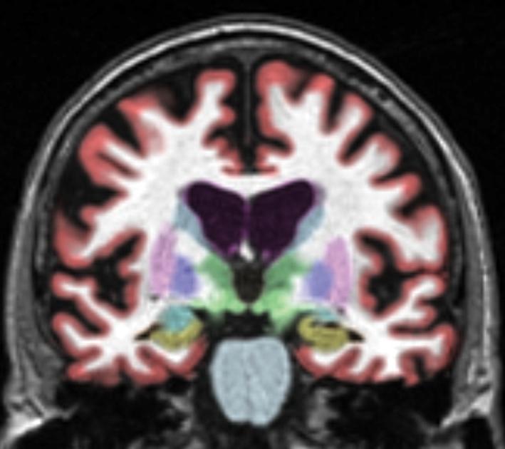
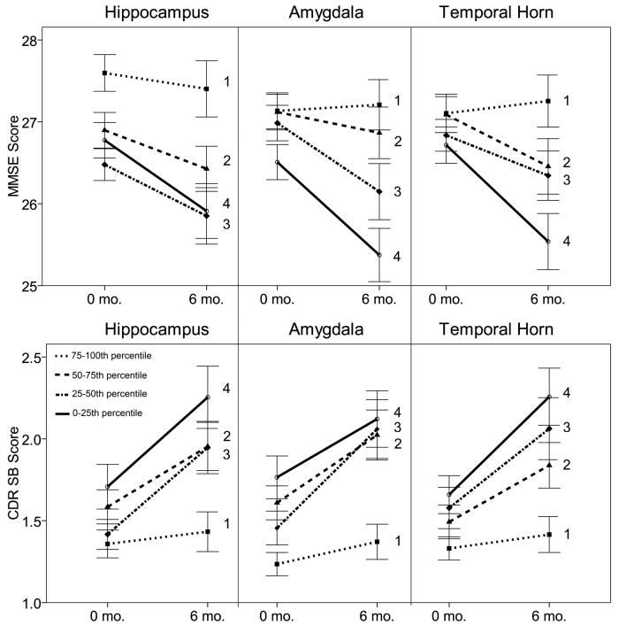
Medial temporal lobe (MTL) atrophy is associated with increased risk for conversion to Alzheimer disease, but manual tracing techniques and even semiautomated techniques for volumetric assessment are not practical in the clinical setting. In addition, most studies that examined MTL atrophy in Alzheimer disease have focused only on the hippocampus. It is unknown the extent to which volumes of amygdala and temporal horn of the lateral ventricle predict subsequent clinical decline. This study examined whether measures of hippocampus, amygdala, and temporal horn volume predict clinical decline over the following 6-month period in patients with mild cognitive impairment (MCI). Fully automated volume measurements were performed in 269 MCI patients. Baseline volumes of the hippocampus, amygdala, and temporal horn were evaluated as predictors of change in Mini-mental State Examination and Clinical Dementia Rating Sum of Boxes over a 6-month interval. Fully automated measurements of baseline hippocampus and amygdala volumes correlated with baseline delayed recall scores. Patients with smaller baseline volumes of the hippocampus and amygdala or larger baseline volumes of the temporal horn had more rapid subsequent clinical decline on Mini-mental State Examination and Clinical Dementia Rating Sum of Boxes. Fully automated and rapid measurement of segmental MTL volumes may help clinicians predict clinical decline in MCI patients.
Figures
References
-
- Petersen RC, Smith GE, Waring SC, et al. Mild cognitive impairment: clinical characterization and outcome. Arch Neurol. 1999;56:303–308. - PubMed
-
- Barnes J, Boyes RG, Lewis EB, et al. Automatic calculation of hippocampal atrophy rates using a hippocampal template and the boundary shift integral. Neurobiol Aging. 2007;28:1657–1663. - PubMed
-
- Crum WR, Scahill RI, Fox NC. Automated hippocampal segmentation by regional fluid registration of serial MRI: validation and application in Alzheimer's disease. Neuroimage. 2001;13:847–855. - PubMed
-
- Folstein MF, Folstein SE, McHugh PR. Mini-mental state: a practical method for grading the cognitive state of patients for the clinician. J Psychiatr Res. 1975;12:189–198. - PubMed
Publication types
MeSH terms
Grants and funding
LinkOut - more resources
Full Text Sources
Medical
Molecular Biology Databases
