. 2009 Aug;16(8):953-62.
doi: 10.1038/gt.2009.59. Epub 2009 May 28.

A muscle-targeting peptide displayed on AAV2 improves muscle tropism on systemic delivery

Affiliations

A muscle-targeting peptide displayed on AAV2 improves muscle tropism on systemic delivery

C-Y Yu et al. Gene Ther. 2009 Aug.

Abstract

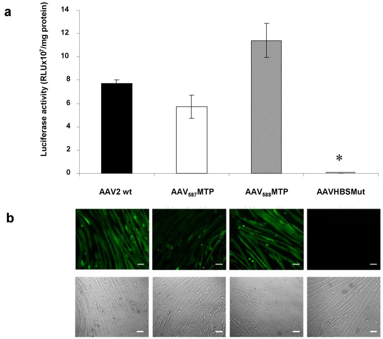
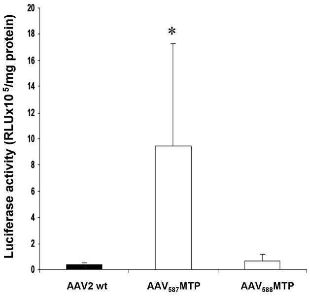
Adeno-associated virus (AAV) has become a leading gene transfer vector for striated muscles. However, the AAV vectors also exhibit broad tropisms after systemic delivery. In an attempt to improve muscle tropism, we inserted a 7-amino-acid (ASSLNIA) muscle-targeting peptide (MTP) in the capsids of AAV2 at residue 587 or 588, generating AAV(587)MTP and AAV(588)MTP. In vitro studies showed that both viruses diminished their infectivity on non-muscle cell lines as well as on un-differentiated myoblasts; however, preserved or enhanced their infectivity on differentiated myotubes. AAV(587)MTP, but not AAV(588)MTP, also abolished its heparin-binding capacity and infected myotubes in a heparin-independent manner. Furthermore, in vivo studies by intravenous vector administration in mice showed that AAV(587)MTP enhanced its tropism to various muscles and particularly to the heart (24.3-fold of unmodified AAV2), whereas reduced its tropism to the non-muscle tissues such as the liver, lungs, spleen and so on. This alteration of tissue tropism is not simply because of the loss of heparin-binding, as a mutant AAV2 (AAVHBSMut) containing heparin-binding site mutations lost infectivity on both non-muscle and muscle cells. Furthermore, free MTP peptide, but not the scrambled control peptide, competitively inhibited AAV(587)MTP infection on myotubes. These results suggest that AAV2 could be re-targeted to the striated muscles by a MTP inserted after residue 587 of the capsids. This proof of principle study showed first evidence of peptide-directed muscle targeting on systemic administration of AAV vectors.


Figures

Figure 1
Figure 1. Construction of AAV mutants
(a) Schematic representation of modified AAV2 capsid amino acid sequences. The peptide encoding ASSLNIA amino acid sequence flanked by two different linkers was inserted after residue 587 or 588 in the AAV2 capsid. The amino acid changes in AAVHBSMut capsid compared to the wild-type AAV2 are indicated. (b) Capsid protein analysis of modified AAV vectors by Western blotting. Similar numbers of AAV genome-containing particles (2×10¹⁰) were separated on 10% SDS-PAGE and analyzed by Western blotting, using anti-AAV2 capsid guinea pig sera.
Figure 2
Figure 2. Efficiency of modified AAV-mediated gene transfer to targeted C2C12 myotubes
(a) Murine C2C12 myotubes were infected with 2×10¹⁰ genomic particles/per well of AAV-CMV-Luc vector which carries either unmodified capsid, peptide-inserted capsid, or heparin-binding mutated capsid in 24-well plates. After 3 days, myotubes were replaced by fresh DMEM containing 2% horse serum and subsequently incubated for 6 days. Luciferase activity was then analyzed to evaluate the transduction efficiencies of modified AAV vectors. Data are shown as a bar graph with mean±standard error of the mean (SEM). *P<0.05 vs. unmodified AAV2 vector. (b) C2C12 myotubes were next transduced with AAV-CB-EGFP vectors at 1×10¹⁰ genomic particles/per well in 24-well plates. EGFP expression driven by the CB promoter was then observed under a Nikon TE-300 inverted fluorescent microscope. Pictures were taken at 72 hours after infection. Fluorescent photography is shown in the upper panel and the morphology of C2C12 myotubes on the same field as the fluorescent image is displayed in the lower panel. Scale bar, 100μm.
Figure 3
Figure 3. Analysis of mutant capsid virus binding to heparin
(a,b) Heparin-affinity column analysis. 5×10¹¹ of unmodified or peptide-inserted viruses were loaded onto a prepacked and equilibrated 1 ml heparin column. Viral particles appeared in the flow-through, wash, and elution fractions were then detected by DNA dot-blot with CMV probe. The fractions from the heparin-affinity column analysis were also analyzed by Western blot using guinea pig anti-AAV2 serum. The positions of VP1, VP2, and VP3 are indicated. I: Input; F: Flow-through; W: Wash step; E: Elution. (c) Evaluation of HSPG-dependent AAV transduction in C2C12 myotubes. C2C12 myotubes were infected with AAV-CMV-Luc vectors carrying unmodified or peptide-inserted capsids in the absence or presence of 30 μg/ml heparin and analyzed for luciferase expression to examine the HSPG dependence of vectors. Data are shown as mean±SEM. *Indicates P<0.05 vs. transduction in the absence of heparin. (d) Competitive blocking experiment by synthesized MTP in C2C12 myotubes. C2C12 myotubes were infected with AAV-CMV-Luc vectors in the presence or absence of synthesized free peptides. Level of gene transduction efficiency of peptide-modified vectors and unmodified AAV virus were compared by evaluating luciferase expression. Data are mean values±SEM. *P<0.05 vs. value in the absence of peptide.
Figure 4
Figure 4. Intramuscular delivery (i.m.) of peptide-modified AAV vectors in mice
Luciferase activities were obtained from the TA muscles of mice injected with 2.5×10¹⁰ genomic particles of AAV-CMV-Luc vector carrying wild-type or modified capsids 4 weeks before examination (n=4 TA muscles for each vector tested). Data are expressed as mean±SEM. * P<0.05 vs. unmodified AAV2 vector.
Figure 5
Figure 5. In vivo AAV-mediated gene transduction after intravenous delivery
9×10¹¹ genomic particles of AAV were delivered to 8-week old male mice via tail vein injection (n=5 for unmodified AAV2, n=6 for AAV587MTP, and n=5 for AAV588MTP vector). Luciferase reporter gene expression in major organs was analyzed one month after delivery. (a) Luciferase activities in cardiac muscle after systemic delivery of peptide-modified vectors. (b) Luciferase activities in striated muscles after systemic AAV administration. (c) Luciferase activities in non-muscle organs after intravenous injection of AAV vectors. *P<0.05 vs. unmodified AAV2 vector. Results are expressed as mean±SEM.
Figure 6
Figure 6. In vivo vector distribution after intravenous delivery
9×10¹¹ viral particles of AAV were administered to 2-month old male mice via tail vein injection (n=5 for unmodified AAV2, and n=6 for AAV587MTP). Vector distribution was quantified by real-time PCR. (a) AAV genome distribution in non-muscle and muscle tissues. (b) Quantify hepatic AAV viral genomes after systemic delivery. Data represent means±SEM. * P<0.05 vs. unmodified AAV2 vector.

Similar articles

Cited by

References

    1. Cox GA, Cole NM, Matsumura K, Phelps SF, Hauschka SD, Campbell KP, et al. Overexpression of dystrophin in transgenic mdx mice eliminates dystrophic symptoms without toxicity. Nature. 1993;364:725–729. - PubMed
    1. Amalfitano A, McVie-Wylie AJ, Hu H, Dawson TL, Raben N, Plotz P, et al. Systemic correction of the muscle disorder glycogen storage disease type II after hepatic targeting of a modified adenovirus vector encoding human acid-alpha-glucosidase. Proc Natl Acad Sci U S A. 1999;96:8861–8866. - PMC - PubMed
    1. Kessler PD, Podsakoff GM, Chen X, McQuiston SA, Colosi PC, Matelis LA, et al. Gene delivery to skeletal muscle results in sustained expression and systemic delivery of a therapeutic protein. Proc Natl Acad Sci U S A. 1996;93:14082–14087. - PMC - PubMed
    1. Greelish JP, Su LT, Lankford EB, Burkman JM, Chen H, Konig SK, et al. Stable restoration of the sarcoglycan complex in dystrophic muscle perfused with histamine and a recombinant adeno-associated viral vector. Nat Med. 1999;5:439–443. - PubMed
    1. Wolff JA, Malone RW, Williams P, Chong W, Acsadi G, Jani A, et al. Direct gene transfer into mouse muscle in vivo. Science. 1990;247:1465–1468. - PubMed

Publication types