

Overexpression of insulin receptor substrate-1 and hepatitis Bx genes causes premalignant alterations in the liver
- PMID: 19475691
- PMCID: PMC2754284
- DOI: 10.1002/hep.22856
Overexpression of insulin receptor substrate-1 and hepatitis Bx genes causes premalignant alterations in the liver
Abstract
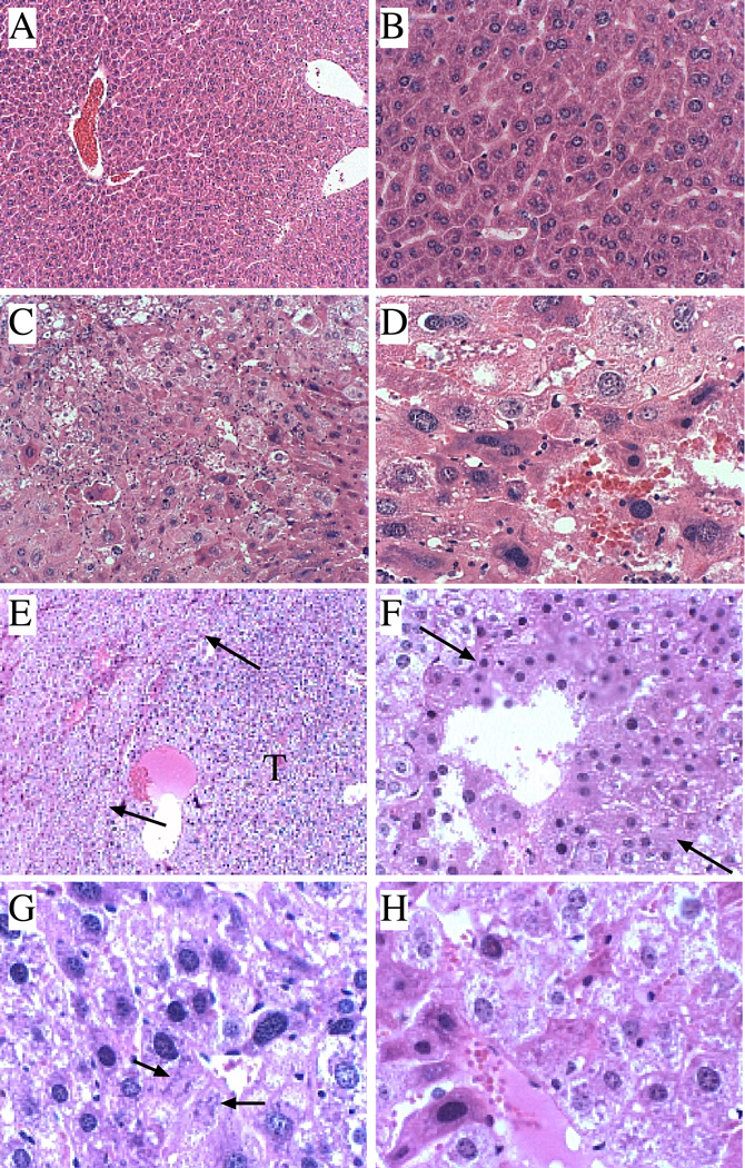
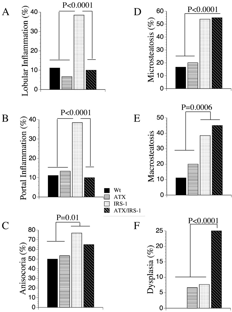
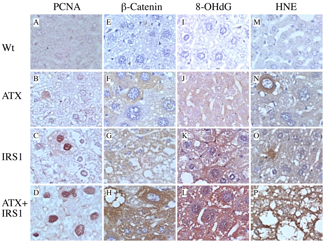
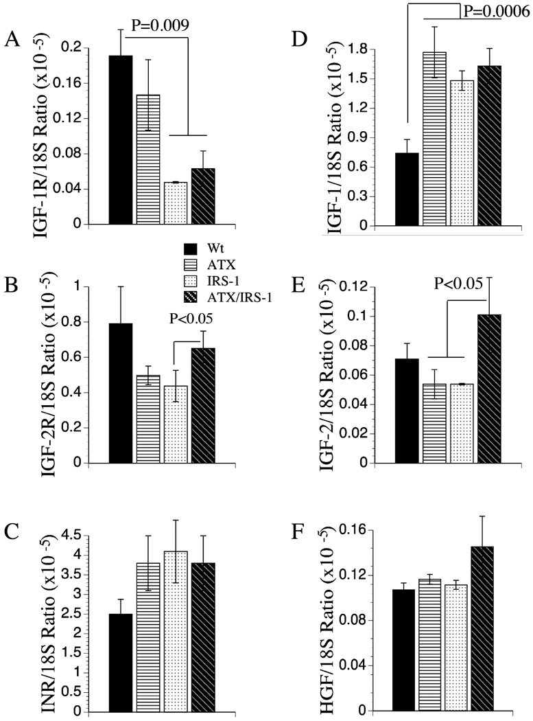
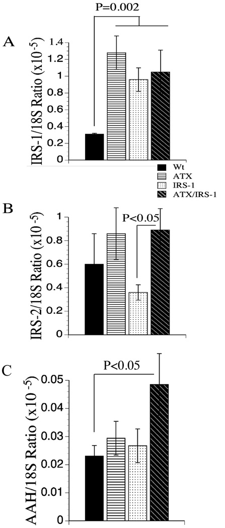
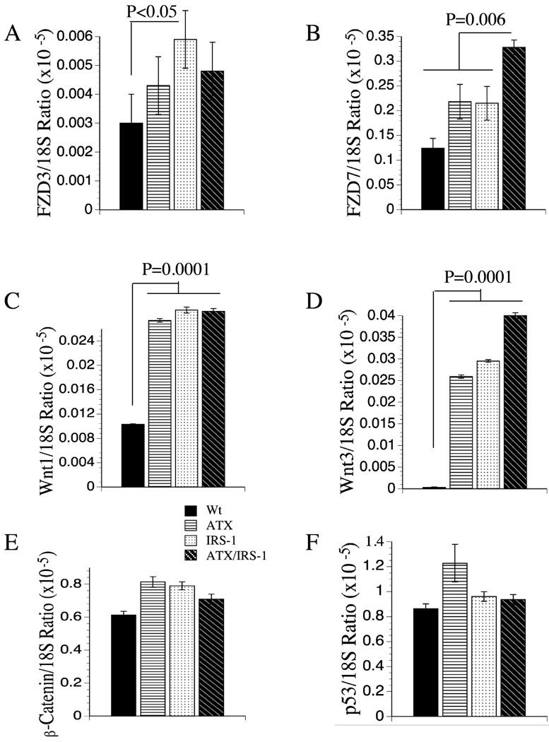
Activation of the insulin (IN)/insulin receptor substrate-1 (IRS-1)/mitogen-associated protein kinase (MAPK) and the Wnt/beta-catenin signaling cascades occurs frequently in hepatocellular carcinoma (HCC) associated with persistent viral infection. The aims of this study were to provide a chronic proliferative stimulus through IRS-1 in the context of hepatitis Bx (HBx) protein expression in transgenic mice and determine if constitutive expression of these genes is sufficient to cause hepatocyte dysplasia and cellular transformation. We generated transgenic mice in which the HBx (ATX), IRS-1, or both (ATX+/IRS-1) genes were expressed under a liver-specific promoter. We also assessed histology and oxidative damage as well as up-regulation of molecules related to these signal transduction cascades in the liver by quantitative reverse-transcriptase polymerase chain reaction. Whereas mice with a single transgene (ATX or IRS-1) did not develop tumors, ATX+/IRS-1+ double transgenic livers had increased frequency of hepatocellular dysplasia and developed HCC. All three transgenic lines had significantly increased insulin growth factor 1 (IGF-1), Wnt 1 and Wnt 3 mRNA levels, and evidence of DNA damage and oxidative stress. The ATX+/IRS+ double transgenic mice were distinguished by having the highest level of activation of Wnt 3 and Frizzled 7 and selectively increased expression of IGF-II, proliferating cell nuclear antigen, and aspartyl-(asparaginyl)-beta-hydroxylase, a gene associated with increased cell migration.
Conclusion: These results suggest that continued expression of the ATX or IRS-1 transgenes can contribute to hepatocyte transformation but are not sufficient to trigger neoplastic changes in the liver. However, dual expression that activates both the IN/IRS-1/MAPK and Wnt/beta-catenin cascades is sufficient to cause dysplasia and HCC in a previously normal liver.
Conflict of interest statement
There is no conflict of interest to disclose.
Figures
References
-
- Parkin DM, Bray F, Ferlay J, Pisani P. Global cancer statistics, 2002. CA Cancer J Clin. 2005;55:74–108. - PubMed
-
- Block TM, Mehta AS, Fimmel CJ, Jordan R. Molecular viral oncology of hepatocellular carcinoma. Oncogene. 2003;22:5093–5107. - PubMed
-
- El-Serag HB, Rudolph KL. Hepatocellular carcinoma: epidemiology and molecular carcinogenesis. Gastroenterology. 2007;132:2557–2576. - PubMed
-
- Breuhahn K, Vreden S, Haddad R, Beckebaum S, Stippel D, Flemming P, Nussbaum T, et al. Molecular profiling of human hepatocellular carcinoma defines mutually exclusive interferon regulation and insulin-like growth factor II overexpression. Cancer Res. 2004;64:6058–6064. - PubMed
-
- Sohda T, Yun K, Iwata K, Soejima H, Okumura M. Increased expression of insulin-like growth factor 2 in hepatocellular carcinoma is primarily regulated at the transcriptional level. Lab Invest. 1996;75:307–311. - PubMed
Publication types
MeSH terms
Substances
Grants and funding
- CA123544/CA/NCI NIH HHS/United States
- AA-12908/AA/NIAAA NIH HHS/United States
- R56 CA095388/CA/NCI NIH HHS/United States
- R01 CA123544/CA/NCI NIH HHS/United States
- AA-11431/AA/NIAAA NIH HHS/United States
- R56 AA011431/AA/NIAAA NIH HHS/United States
- AA-02666/AA/NIAAA NIH HHS/United States
- CA035711/CA/NCI NIH HHS/United States
- R01 AA011431/AA/NIAAA NIH HHS/United States
- R37 AA002666/AA/NIAAA NIH HHS/United States
- R01 CA095388/CA/NCI NIH HHS/United States
- R37 AA011431/AA/NIAAA NIH HHS/United States
- CA095388/CA/NCI NIH HHS/United States
- R01 AA012908/AA/NIAAA NIH HHS/United States
- K24 AA016126/AA/NIAAA NIH HHS/United States
- R01 AA002666/AA/NIAAA NIH HHS/United States
- R01 CA035711/CA/NCI NIH HHS/United States
- R37 CA035711/CA/NCI NIH HHS/United States
LinkOut - more resources
Full Text Sources
Medical
Miscellaneous