. 2009 Jun;127(2):279-88.
doi: 10.1111/j.1365-2567.2008.03016.x.

Mannan-binding lectin deficiency modulates the humoral immune response dependent on the genetic environment

Affiliations

Mannan-binding lectin deficiency modulates the humoral immune response dependent on the genetic environment

Marieta Ruseva et al. Immunology. 2009 Jun.

Abstract

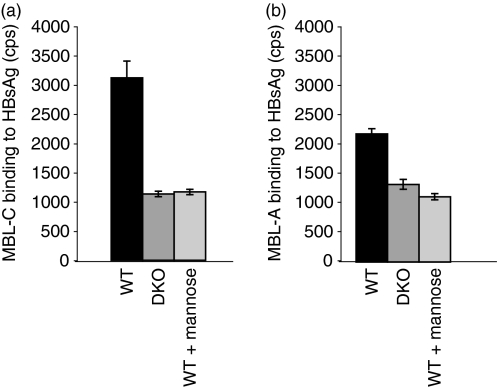
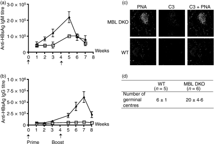
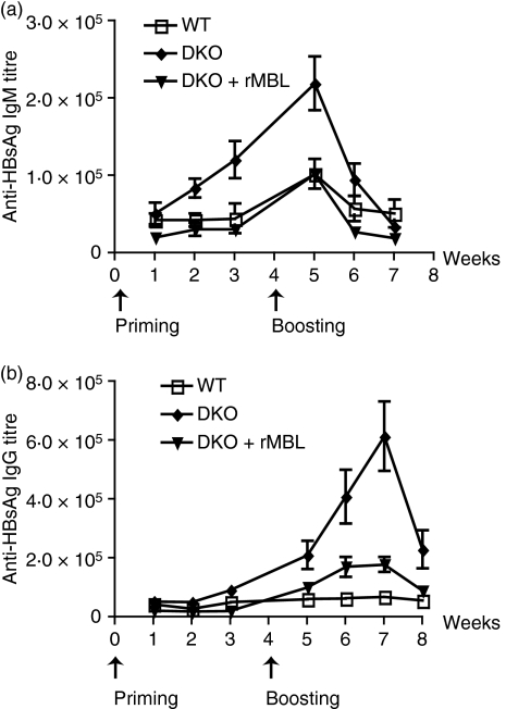
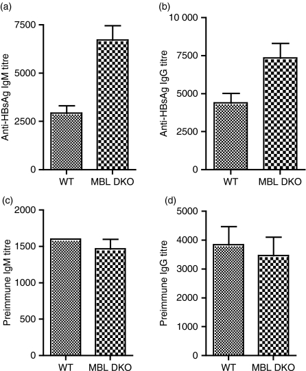
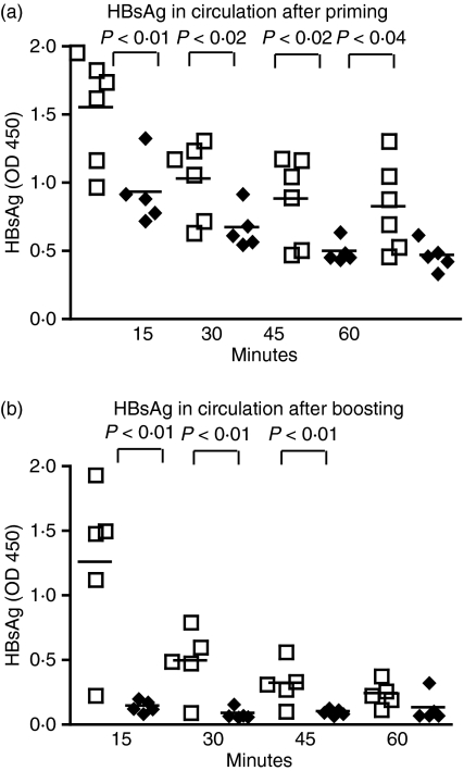
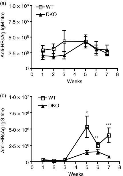
Mannan-binding lectin (MBL) is a plasma protein implicated in innate immune defence against a broad range of microorganisms, including viruses. It is also thought that MBL plays a role in the recruitment of the specific clonal immune response. This was studied by injecting soluble hepatitis B surface antigen (HBsAg) intravenously into mice deficient in both MBL-A and MBL-C (MBL DKO mice). The MBL DKO animals on mixed genetic background (SV129EvSv x C57BL/6) produced higher antibody titres than the wild-type littermates. After primary challenge with the antigen the immunoglobulin M anti-HBsAg antibody titres were threefold higher in the MBL DKO mice than in the wild-type mice. Following the boost, the immunoglobulin G anti-HBsAg antibody titres were 10-fold higher in the MBL DKO mice, suggesting that MBL plays a role in a negative feedback regulation of adaptive immunity. However, the modulating effect of MBL was dependent on the genetic environment. The MBL DKO mice backcrossed on a C57BL/6 background showed the opposite response with the MBL DKO mice now producing fewer antibodies than the wild-type animals, whereas MBL deficiency in mice with the SV129EvSv background did not show any effect in antibody production. These findings indicate that the modifying effect of MBL on the humoral immune response is influenced by the genetic environment.


Figures

Figure 1
Figure 1
Mannan-binding lectin A (MBL-A) and MBL-C bind specifically to hepatitis B surface antigen (HBsAg). MBL-A and MBL-C binding to HBsAg was analysed in a time-resolved immunofluorometry assay, where HBsAg were coated on an enzyme-linked immunsorbent assay plate and then exposed to sera from WT and MBL DKO animals, followed by development with specific anti-mouse MBL-A and MBL-C antibodies. The sera were diluted in CaCl2-containing buffer to enable the MBL interaction with ligands or inhibited by mannose-containing buffer. The y-axis represents counts per second (cps) measured in the wells.
Figure 2
Figure 2
Antibody titres against hepatitis B surface antigen (HBsAg) are elevated in mannose-binding lectin double knockout (MBL DKO; 129 SvEv × C57BL/6) mice compared with wild-type (WT) control littermates. MBL DKO and WT mice were challenged with 8 μg HBsAg intravenously (a and b) The antigen-specific immunoglobulin M (IgM) and IgG titres were determined by time-resolved immunofluorometry and followed for 3 weeks after priming and boost. At least five animals in each group were used. The data shown are representative of two individual experiments. (c) Immunofluorescent detection of C3 deposits within the GC. Spleens were harvested 5 weeks after the boosting. Cryosections were treated with PNA and anti-C3d antibody. Sections are representative of spleens analysed from at least five mice per group. (d) Quantification of the number of germinal centres per splenic section. MBL DKO and WT mice were harvested after the boost at the peak of the IgG response. Splenic sections were stained with PNA and the number of germinal centres was counted per section. At least five animals per group and five individual sections per spleen were analysed.
Figure 3
Figure 3
Recombinant mannose-binding lectin (rMBL) reconstitutes the antibody responses to hepatitis B surface antigen (HBsAg) in MBL double knockout (DKO; 129 SvEv x C57BL/6) mice. MBL DKO, MBL DKO supplemented with rMBL at the time of the antigen challenge and wild-type (WT) mice were immunized with 8 μg HBsAg intravenously (a and b) The antigen-specific IgM and IgG titres were determined by time-resolved immunofluorometry and followed for 3 weeks after priming and boost. At least 10 animals in each group were used.
Figure 4
Figure 4
Preimmune hepatitis B surface antigen (HBsAg)-specific immunoglobulin M (IgM) levels are elevated in the mannose-binding lectin double knockout (MBL DKO) mice. The preimmune HBsAg-specific IgM (a) and IgG (b) titres were determined by a time-resolved immunofluorometry (TRIFMA) assay where microtitre wells were coated with HBsAg and exposed to different dilutions of non-immune sera. At least 10 animals per group were tested. Groups were compared using the Mann–Whitney test for significance. Significant differences were observed for anti-HBsAg IgM titres (P = 0·007) and anti-HBsAg IgG titres (P = 0·03). Preimmune IgM (c) and IgG (d) levels. The preimmune serum levels of IgM and IgG were determined by a TRIFMA assay where microtitre wells were coated with anti-mouse immunoglobulin and exposed to different dilutions of non-immune sera. Groups were compared using the Mann–Whitney test for significance. In both cases, no significant differences were found between the groups (P > 0·05).
Figure 5
Figure 5
Hepatitis B surface antigen (HBsAg) is cleared from circulation more efficiently in mannose-binding lectin double knockout (MBL DKO) mice than in the wild-type (WT) animals. Non-primed (a) and primed (b) MBL DKO and WT animals were challenged with HBsAg intravenously and serum samples were collected at different time-points after the challenge. The levels of HBsAg in circulation were determined by enzyme-linked immunosorbent assay. Individual animals are shown as individual symbols. The different groups were compared by Mann–Whitney test analysis for significance and P-values are noted in the figure.
Figure 6
Figure 6
The hepatitis B surface antigen (HBsAg)-specific immunoglobulin M (IgM) and IgG titres in mannose-binding lectin double knockout (MBL DKO) F6 and C57BL/6 mice. MBL DKO F6 and C57BL/6 mice were challenged with 2 μg HBsAg intravenously. The antigen-specific IgM (a) and IgG (b) titres were determine by time-resolved immunofluorometry and followed for 3 weeks after priming and after boost. At least five animals in each group were used. The data shown are representative of two individual experiments. Groups were compared using the Mann–Whitney test for significance. The stars annotate the time-points at which significant differences in the titres were seen (P > 0·05).

References

    1. Hart ML, Saifuddin M, Spear GT. Glycosylation inhibitors and neuraminidase enhance human immunodeficiency virus type 1 binding and neutralization by mannose-binding lectin. J Gen Virol. 2003;2:353–60. - PubMed
    1. Thielens NM, Tacnet-Delorme P, Arlaud GJ. Interaction of C1q and mannan-binding lectin with viruses. Immunobiology. 2002;205:563–74. - PubMed
    1. Lubinski J, Nagashunmugam T, Friedman HM. Viral interference with antibody and complement. Semin Cell Dev Biol. 1998;9:329–37. - PMC - PubMed
    1. Friedman HM, Wang L, Pangburn MK, Lambris JD, Lubinski J. Novel mechanism of antibody-independent complement neutralization of herpes simplex virus type 1. J Immunol. 2000;165:4528–36. - PubMed
    1. Matsushita M, Fujita T. Activation of the classical complement pathway by mannose-binding protein in association with a novel C1s-like serine protease. J Exp Med. 1992;176:1497–502. - PMC - PubMed

Publication types