

Induction of tyrosine hydroxylase mRNA by nicotine in rat midbrain is inhibited by mifepristone
- PMID: 19476543
- PMCID: PMC2731240
- DOI: 10.1111/j.1471-4159.2009.06056.x
Induction of tyrosine hydroxylase mRNA by nicotine in rat midbrain is inhibited by mifepristone
Abstract
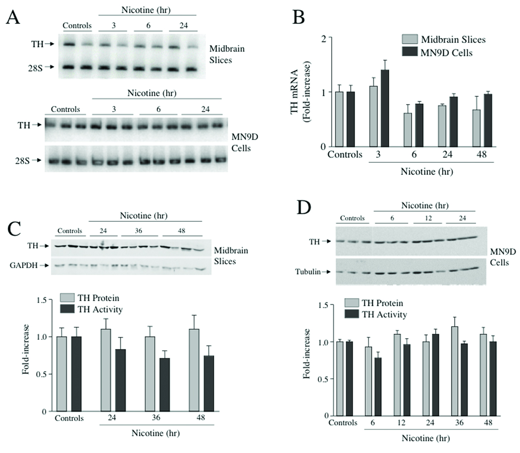
Repeated nicotine administration induces tyrosine hydroxylase (TH) mRNA in rat midbrain. In this study we investigate the mechanisms responsible for this response using two models of midbrain dopamine neurons, rat ventral midbrain slice explant cultures and mouse MN9D cells. In both models nicotine stimulates TH gene transcription rate in a dose-dependent manner. However, this stimulation is short-lived, lasting for 1 h, but less than 3 h, and is not sufficient to induce TH mRNA or TH protein. Nicotine elevates circulating glucocorticoids, which induce TH expression in some model systems. We tested the hypothesis that the effect of nicotine on midbrain TH mRNA is mediated by the glucocorticoid receptor. When rats are administered the glucocorticoid receptor antagonist mifepristone, the induction of TH mRNA by nicotine in both substantia nigra and ventral tegmentum is inhibited. Furthermore, the glucocorticoid receptor agonist dexamethasone stimulates TH gene transcription for sustained periods of time in both midbrain slices and MN9D cells, leading to induction of TH mRNA and TH protein. Our results are consistent with the hypothesis that nicotine induces TH mRNA in midbrain by elevating glucocorticoids, which then act on glucocorticoid receptors in dopamine neurons leading to transcriptional activation of the TH gene.
Figures


Similar articles
-
Nicotinic and muscarinic acetylcholine receptors are essential for the long-term response of tyrosine hydroxylase gene expression to chronic nicotine treatment in rat adrenal medulla.Brain Res Mol Brain Res. 2004 Jul 26;126(2):188-97. doi: 10.1016/j.molbrainres.2004.04.007. Brain Res Mol Brain Res. 2004. PMID: 15249143
-
Adrenal tyrosine hydroxylase activity and gene expression are increased by intraventricular administration of nicotine.J Pharmacol Exp Ther. 2001 Jan;296(1):15-21. J Pharmacol Exp Ther. 2001. PMID: 11123357
-
Activation of tyrosine hydroxylase mRNA translation by cAMP in midbrain dopaminergic neurons.Mol Pharmacol. 2008 Jun;73(6):1816-28. doi: 10.1124/mol.107.043968. Epub 2008 Mar 18. Mol Pharmacol. 2008. PMID: 18349104 Free PMC article.
-
Involvement of alpha7 nicotinic acetylcholine receptors in activation of tyrosine hydroxylase and dopamine beta-hydroxylase gene expression in PC12 cells.J Neurochem. 2000 Nov;75(5):1997-2005. doi: 10.1046/j.1471-4159.2000.0751997.x. J Neurochem. 2000. PMID: 11032889
-
Involvement of alpha 7 nicotinic acetylcholine receptors in gene expression of dopamine biosynthetic enzymes in rat brain.J Pharmacol Exp Ther. 2002 Dec;303(3):896-903. doi: 10.1124/jpet.102.039198. J Pharmacol Exp Ther. 2002. PMID: 12438507
Cited by
-
The Sensory Impact of Nicotine on Noradrenergic and Dopaminergic Neurons of the Nicotine Reward - Addiction Neurocircuitry.J Addict Res Ther. 2016 Apr;7(2):274. doi: 10.4172/2155-6105.1000274. Epub 2016 Apr 7. J Addict Res Ther. 2016. PMID: 27347434 Free PMC article.
-
Paecilomycies japonica reduces repeated nicotine-induced neuronal and behavioral activation in rats.BMC Complement Altern Med. 2015 Jul 14;15:227. doi: 10.1186/s12906-015-0739-8. BMC Complement Altern Med. 2015. PMID: 26169054 Free PMC article.
-
Enhanced synthesis and release of dopamine in transgenic mice with gain-of-function α6* nAChRs.J Neurochem. 2014 Apr;129(2):315-27. doi: 10.1111/jnc.12616. Epub 2013 Dec 13. J Neurochem. 2014. PMID: 24266758 Free PMC article.
-
A critical period of vulnerability to adolescent stress: epigenetic mediators in mesocortical dopaminergic neurons.Hum Mol Genet. 2016 Apr 1;25(7):1370-81. doi: 10.1093/hmg/ddw019. Epub 2016 Jan 28. Hum Mol Genet. 2016. PMID: 26908623 Free PMC article.
-
Programming of Dopaminergic Neurons by Neonatal Sex Hormone Exposure: Effects on Dopamine Content and Tyrosine Hydroxylase Expression in Adult Male Rats.Neural Plast. 2016;2016:4569785. doi: 10.1155/2016/4569785. Epub 2016 Jan 10. Neural Plast. 2016. PMID: 26904299 Free PMC article.
References
-
- Bowyer JF, Frame LT, Clausing P, Nagamoto-Combs K, Osterhout CA, Sterling CR, Tank AW. The long-term effects of amphetamine neurotoxicity on tyrosine hydroxylase mRNA and protein in aged rats. J Pharmacol Exp Thera. 1998;286:1074–1085. - PubMed
-
- Bradford MM. A rapid and sensitive method for the quantitation of microgram quantities of protein utilizing the principle of protein-dye binding. Anal Biochem. 1976;72:248–254. - PubMed
-
- Caggiula AR, Donny EC, Epstein LH, Sved AF, Knopf S, Rose C, McAllister CG, Antelman SM, Perkins KA. The role of corticosteroids in nicotine's physiological and behavioral effects. Psychoneuroendocrinology. 1998;23:143–159. - PubMed
Publication types
MeSH terms
Substances
Grants and funding
LinkOut - more resources
Full Text Sources
