. 2009 May 29:9:31.
doi: 10.1186/1471-213X-9-31.

Tbx1 and Brn4 regulate retinoic acid metabolic genes during cochlear morphogenesis

Affiliations

Tbx1 and Brn4 regulate retinoic acid metabolic genes during cochlear morphogenesis

Evan M Braunstein et al. BMC Dev Biol. .

Abstract

Background: In vertebrates, the inner ear is comprised of the cochlea and vestibular system, which develop from the otic vesicle. This process is regulated via inductive interactions from surrounding tissues. Tbx1, the gene responsible for velo-cardio-facial syndrome/DiGeorge syndrome in humans, is required for ear development in mice. Tbx1 is expressed in the otic epithelium and adjacent periotic mesenchyme (POM), and both of these domains are required for inner ear formation. To study the function of Tbx1 in the POM, we have conditionally inactivated Tbx1 in the mesoderm while keeping expression in the otic vesicle intact.

Results: Conditional mutants (TCre-KO) displayed malformed inner ears, including a hypoplastic otic vesicle and a severely shortened cochlear duct, indicating that Tbx1 expression in the POM is necessary for proper inner ear formation. Expression of the mesenchyme marker Brn4 was also lost in the TCre-KO. Brn4-;Tbx1+/-embryos displayed defects in growth of the distal cochlea. To identify a potential signal from the POM to the otic epithelium, expression of retinoic acid (RA) catabolizing genes was examined in both mutants. Cyp26a1 expression was altered in the TCre-KO, while Cyp26c1 showed reduced expression in both TCre-KO and Brn4-;Tbx1+/- embryos.

Conclusion: These results indicate that Tbx1 expression in the POM regulates cochlear outgrowth potentially via control of local retinoic acid activity.


Figures

Figure 1
Figure 1
Inner ear defects in the TCre-KO. At E15.5, a paint-filled inner ear of a control embryo (A) displays a cochlea (c) consisting of 1.75 turns and a vestibular system consisting of the saccule (s), utricle (u), endolymphatic duct (ed) and lateral, posterior and superior semicircular canals (lsc, psc, ssc). A transverse section though the inner ear of an E17.5 control embryo (B) displays the sensory structures of the ear with an intact organ of Corti (bracketed in inset) and the surrounding capsule. Bone and cartilage staining of an E17.5 control embryo (C) reveals the vestibular capsule (vc) and cochlear capsule (cc). Paint-fill of the inner ear of an E15.5 TCre-KO embryo (D) reveals malformations of both the auditory and vestibular system. The cochlea is severely shortened, the vestibular structures are hypoplastic and the endolymphatic duct (which did not completely fill with paint in (A)) is enlarged. At E17.5, the inner ears of TCre-KO embryos (E) are malformed and display only the hook region of the cochlea (c) with no recognizable organ of Corti (inset), and hypoplastic semicircular canals (lsc, ssc). Bone and cartilage staining (F) reveals a malformed cochlear capsule (cc), while the vestibular capsule (vc) appears relatively unaffected. Scale bar in B is 200 μm.
Figure 2
Figure 2
Otic markers and vesicle morphology are unaffected at E10.5 in TCre-KO mutants. Expression of sensory (A-F) and neurogenic (G-J) markers are unchanged in TCre-KO embryos compared to controls at E10.5. Ventral markers Pax2 and Otx1, as well as dorsal markers Bmp4 and Dlx5 are normal in TCre-KO mutants (A'-D'). Gata3 expression in the lateral otic vesicle is also unchanged in the TCre-KO, visualized in cross sections through the otic vesicle (F'). Expression of anterior markers NeuroD, Fgf3, Lnfg and Fgf10 display normal expression in the CVG in both mutants (G'-J') and controls (G-J). Expression of Lmx1a, which marks the growing endolymphatic duct, is also unchanged (K, K'). Microdissected otic vesicles are shown at E10.5 (L, L') and E11.5 (M, M'). At E10.5, the morphology of the otic vesicle in control (L) and TCre-KO (L') embryos is similar. By E11.5, the otic vesicles of TCre-KO embryos (M') are malformed and hypoplastic compared to controls (M). Scale bar in L is 250 μm and applies to all panels except (E, E') and (F, F'). Scale bars in E' and F' are 200 μm.
Figure 3
Figure 3
TCre-KO embryos exhibit defects in cell proliferation and survival. Transverse sections at E11.5 of control (A) and TCre-KO (C) embryos labeled for mitotic cells using an Ab against phospho-Histone 3. TCre-KO embryos display a decrease in cell proliferation in the otic epithelium compared to controls upon quantification (E). Proliferation was reduced in both dorsal and ventral regions of the otic vesicle, but not in the POM. For the dorsal otic vesicle, 11.2 and 21.4 positive cells were observed for mutant and control, respectively, while 14.0 and 22.4 positive cells were observed observed for the ventral otic vesicle. TCre-KO embryos exhibit an increase in apoptosis at E11.5, identified by an Ab to active Caspase 3 (B, D). Arrow and bracketed area in (d) mark apoptotic cells. Quantification of apoptotic cells in control and mutant otic epithelium is shown (F). Asterisks indicate significant difference (* = p < 0.05, ** = p < 0.01, one-tailed, unpaired Student's t-test). Error bars represent 95% confidence interval. Boxed areas are magnified in a-d. Scale bar indicates 200 μm in (A), and 50 μm in (a).
Figure 4
Figure 4
Brn4 expression is reduced in the POM of TCre-KO embryos, and Brn4-;Tbx1+/- embryo display defects in cochlea coiling. Sagittal sections of control (A) and TCre-KO (B) embryos at E10.5 stained with an Ab against Brn4. Expression in the POM is lost in TCre-KO embryos, however neural tube (nt) expression is unaffected. Brn4-;Tbx1+/- embryos display a shortened cochlear duct. Lateral views of paint-filled membranous labyrinths at E15.5–E16.0 of Tbx1+/- (E), Brn4+/-;Tbx1+/- (F), Brn4-(G, H), Brn4-;Tbx1+/- (I, J). The fully developed mouse cochlea normally completes 1.75 turns, and all inner ears of Tbx1+/- mutants and Brn4+/-;Tbx1+/- exhibited normal cochlear growth (E, F). The majority of Brn4- mutant inner ears also displayed a normal cochlea (G), while 20% displayed a slightly shortened cochlea (H). All inner ears Brn4-;Tbx1+/- mutants exhibited a malformed cochlea that failed to form a second turn and instead displayed abnormal twisting or bifurcation (I, J). Arrows point to the cochlear duct in H-J. The endolymphatic sac in E, F, G and J was present but did not fill with paint. cvg, cochleovestibular ganglion; ov, otic vesicle; nt, neural tube; pom, periotic mesenchyme. Scale bar in D is 200 μm, scale bar in F is 350 μm.
Figure 5
Figure 5
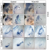
Expression of Cyp26 genes is altered in TCre-KO embryos. Cyp26a1 and Cyp26c1 expression detected by whole mount in situ hybridization at E10.5 in control and TCre-KO embryos (A-H). The plane of section in B, D, F and H is marked in A, C, E, and G, respectively. Cyp26a1 expression in the lateral POM (arrows in B) is absent in TCre-KO mutants (arrows in D), and appears shifted to a more ventral position (compare A an C). Cyp26c1 expression is strongly reduced in the mesenchyme of TCre-KO mutants (compare F and H), but 1st arch expression is unaffected (asterisk in E and G). At E11.5, Cyp26a1 expression remains shifted medially in TCre-KO mutants compared to controls (I-L), especially in posterior sections through the growing cochlear duct (arrows in J and L). Cyp26c1 is strongly reduced in the ventral POM at this stage (M-P), but remains present in the otic epithelium (arrows in M and O). A dotted circle outlines the otic vesicle (ov) in A, C, E, G. Scale bar in P is 200 μm and applies to all panels except A, C, E, G.
Figure 6
Figure 6
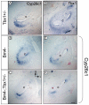
Cyp26c1 expression is reduced in Brn4-;Tbx1+/- embryos. Transverse sections through the cochlear duct (c) probed for Cyp26c1 (A-C, E-F) or Tbx1 (D) in Tbx1+/-, Brn4- and Brn4-;Tbx1+/- embryos at E12.5. Cyp26c1 expression is reduced in the ventromedial POM surrounding the cochlear duct of Brn4-;Tbx1+/- embryos (C and F) compared to Tbx1+/- (A) and Brn4- (B and E) embryos. Tbx1 is expressed in this same region (D) and overlaps with Cyp26c1 expression. Expression of Cyp26c1 in the otic epithelium is unaffected in Brn4-;Tbx1+/- embryos. Scale bar is 200 μm.
Figure 7
Figure 7
Model of Tbx1 and Brn4 function in the POM. Sonic hedgehog (Shh) secreted from the notochord (circle) and floor plate regulates both Tbx1 and Brn4 in the POM (blue). This signal is likely mediated by Gli genes expressed in the POM. Tbx1 and Brn4 activate members of the Cyp26 family also expressed in the POM. These genes catabolize active retinoic acid (RA) secreted from the otic vesicle (green), thereby regulating RA activity in the POM. Brn4 regulates expression of Cyp26c1, while Tbx1 regulates both Cyp26a1 and Cyp26c1. Strict control of RA dosage in the POM is likely crucial for proper cochlear coiling. CD, cochlear duct; ED, endolymphatic duct; POM, periotic mesenchyme.

Similar articles

Cited by

References

    1. Barald KF, Kelley MW. From placode to polarization: new tunes in inner ear development. Development. 2004;131:4119–4130. doi: 10.1242/dev.01339. - DOI - PubMed
    1. Fekete DM, Wu DK. Revisiting cell fate specification in the inner ear. Curr Opin Neurobiol. 2002;12:35–42. doi: 10.1016/S0959-4388(02)00287-8. - DOI - PubMed
    1. Giraldez F, Fritzsch B. The molecular biology of ear development – "Twenty years are nothing". Int J Dev Biol. 2007;51:429–438. doi: 10.1387/ijdb.072390fg. - DOI - PMC - PubMed
    1. Riccomagno MM, Martinu L, Mulheisen M, Wu DK, Epstein DJ. Specification of the mammalian cochlea is dependent on Sonic hedgehog. Genes Dev. 2002;16:2365–2378. doi: 10.1101/gad.1013302. - DOI - PMC - PubMed
    1. Riccomagno MM, Takada S, Epstein DJ. Wnt-dependent regulation of inner ear morphogenesis is balanced by the opposing and supporting roles of Shh. Genes Dev. 2005;19:1612–1623. doi: 10.1101/gad.1303905. - DOI - PMC - PubMed

Publication types

MeSH terms