

Multisystemic eosinophilia resembling hypereosinophilic syndrome in a colony-bred owl monkey (Aotus vociferans)
- PMID: 19476722
- PMCID: PMC2696836
Multisystemic eosinophilia resembling hypereosinophilic syndrome in a colony-bred owl monkey (Aotus vociferans)
Abstract
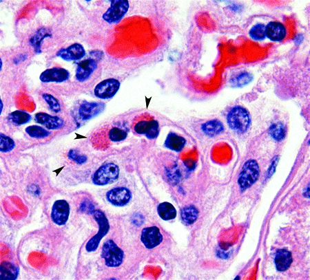
In animals, multisystemic eosinophilic disease is a rare condition characterized by eosinophilic and lymphoplasmacytic infiltrates in various organs. This disorder resembles the human disease known as hypereosinophilic syndrome, a condition defined by prolonged peripheral eosinophilia in the absence of recognizable etiology and associated with end-organ damage. In this report we describe a research-naïve, colony-born, juvenile female owl monkey (Aotus vociferans) who presented clinically with severe respiratory distress and histologically with multiple end-organ infiltration with phenotypically mature eosinophils, plasma cells, and lymphocytes. No tumors or infectious agents were noted either macroscopically or microscopically. Cultures from lung samples revealed no bacteria or fungi. Histologic examination of lung, heart, thymus, liver, spleen, kidney, adrenal, pancreas, stomach, small intestine, and colon revealed no migrating nematode larvae, other parasites, or foreign material that might trigger eosinophilia, nor was there any evidence of or history consistent with an allergic etiology. Given that we ruled out most exogenous and endogenous triggers of eosinophilia, the signs, symptoms, and pathologic findings support the diagnosis of multisystemic eosinophilic disease. To our knowledge, this report is the first description of presumptive hypereosinophilic syndrome in a nonhuman primate.
Figures


References
-
- Albert DL, Brodie SJ, Sasseville VG, Ringler DJ. 1996. Peripheral blood eosinophilia in owl monkeys is associated with increased eosinophilopoeisis yet depressed recruitment kinetics to the Chemokine RANTES. Blood 88:1718–1724 - PubMed
-
- Aroch I, Perl S, Markovics A. 2001. Disseminated eosinophilic disease resembling idiopathic hypereosinophilic syndrome in a dog. Vet Rec 149:386–389 - PubMed
-
- Blomme EA, Foy SH, Chappell KH, La Perle KM. 1999. Hypereosinophilic syndrome with Hodgkin's-like lymphoma in a ferret. J Comp Pathol 120:211–217 - PubMed
-
- Cools J, DeAngelo DJ, Gotlib J, Stover EH, Legare RD, Cortes J, Kutok J, Clark J, Galinsky I, Griffin JD, Cross NC, Tefferi A, Malone J, Alam R, Schrier SL, Schmid J, Rose M, Vandenberghe P, Verhoef G, Boogaerts M, Wlodarska I, Kantarjian H, Marynen P, Coutre SE, Stone R, Gilliland DG. 2003. A tyrosine kinase created by fusion of the PDGFRA and FIP1L1 genes as a therapeutic target of imatinib in idiopathic hypereosinophilic syndrome. N Engl J Med 348:1201–1214 - PubMed
Publication types
MeSH terms
Grants and funding
LinkOut - more resources
Full Text Sources
Medical