Review
. 2009 Jun 6;373(9679):1974-86.
doi: 10.1016/S0140-6736(09)60259-2. Epub 2009 May 22.

Neurofibromatosis type 2

Affiliations
Review

Neurofibromatosis type 2

Ashok R Asthagiri et al. Lancet. .

Abstract

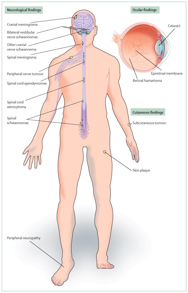
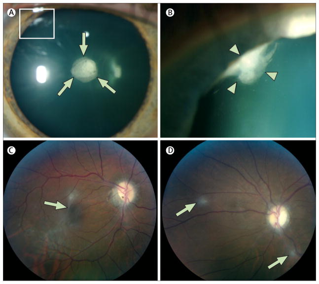
Neurofibromatosis type 2 is an autosomal-dominant multiple neoplasia syndrome that results from mutations in the NF2 tumour suppressor gene located on chromosome 22q. It has a frequency of one in 25,000 livebirths and nearly 100% penetrance by 60 years of age. Half of patients inherit a germline mutation from an affected parent and the remainder acquire a de novo mutation for neurofibromatosis type 2. Patients develop nervous system tumours (schwannomas, meningiomas, ependymomas, astrocytomas, and neurofibromas), peripheral neuropathy, ophthalmological lesions (cataracts, epiretinal membranes, and retinal hamartomas), and cutaneous lesions (skin tumours). Optimum treatment is multidisciplinary because of the complexities associated with management of the multiple, progressive, and protean lesions associated with the disorder. We review the molecular pathogenesis, genetics, clinical findings, and management strategies for neurofibromatosis type 2.


Conflict of interest statement

Conflicts of interest

We declare that we have no conflicts of interest.

Figures

Figure 1
Figure 1
Clinical manifestations of neurofibromatosis type 2
Figure 2
Figure 2. Molecular biology of merlin
(A) Merlin isoform I has three structural regions: a tri-lobed aminoterminal protein 4·1-ezrin-radixin-moesin (FERM) domain, an α helical domain, and a carboxyterminal domain. PKA=cAMP dependent protein kinase A. PAK=p21-activated kinases. MYPT-1-PP1 δ=myosin phosphatase-1 protein phosphatase-1. ser518=serine-518. (B) Active state of merlin. In this state, merlin mediates its tumour suppressor activity through the downstream regulation of various mitogenic intracellular signalling pathways. Most important of these activities is its effects on the phosphoinositide-3 kinase (PI3K)-signalling pathway (PI3K–Akt–MTOR) and the mitogen-activated protein kinase (MAPK) signalling pathway (Ras–Raf–MEK–ERK). EGFR=endothelial growth factor receptor. PIKE-L=phosphatidylinositol 3-kinase enhancer long form. eiF3c=eukaryotic initiation factor 3 subunit c. TRBP=transactivation responsive RNA binding proteins. RalGDS=Ral guanine-nucleotide dissociation stimulator. Rho GTPases=Rho guanosine triphosphatases. N-WASP=Neuronal Wiskott-Aldrich syndrome protein.
Figure 3
Figure 3. MRI and histological features of schwannomas in neurofibromatosis type 2
(A) Axial T1-weighted contrast-enhanced MRI of a man’s brain reveals bilateral vestibular schwannomas (arrows). Intracanlicular extension (arrowheads) of tumours into the internal auditory meatus is clearly delineated. (B) Axial T1-weighted contrast-enhanced MRI of the cervical spine reveals a dumbbell shaped spinal schwannoma (arrows), showing mass effect on adjacent spinal cord (arrowheads) in a young man. (C) Axial T1-weighted contrast-enhanced, fat-suppressed MRI of a woman’s chest shows a schwannoma (arrow) of an intercostal nerve. (D) Haematoxylin and eosin stained section of an excised vestibular schwannoma reveals a spindle-cell lesion, with cells that assume intermixed dense (Antoni A) (a) and loose (Antoni B) (b) arrangements.
Figure 4
Figure 4. MRI features of meningiomas in neurofibromatosis type 2
(A) Axial T1-weighted contrast-enhanced MRI of a woman’s brain show several enhancing intracranial meningiomas (arrows). (B) Sagittal T1-weighted contrast-enhanced MRI of the cervical spine in a young man shows a spinal meningioma (arrows) with classic dural tail sign (green arrowheads). An enhancing intramedullary tumour (probable ependymoma) (black arrowhead) is visible in the adjacent spinal cord.
Figure 5
Figure 5. MRI and histological features of an ependymoma in neurofibromatosis type 2
(A) Sagittal T1-weighted MRI of the cervical spine in a man reveals a subtle swelling (arrows) of the spinal cord at C7, suggesting the presence of an intramedullary lesion. (B) Sagittal T1-weighted contrast-enhanced MRI in the same patient shows the homogeneous pattern of enhancement in a central location within the spinal cord and frequent multiplicity of ependymomas (string of pearls) (arrows). (C) Haematoxylin and eosin stained section of an excised ependymoma reveals a well delineated, moderately cellular tumour with a monomorphic nuclear morphology that is characterised by round to oval nuclei with salt and pepper speckling of the chromatin (arrows). Occasional perivascular pseudorosette (asterisk) and ependymal rosette arrangements are typical histological findings.
Figure 6
Figure 6. Ocular findings in neurofibromatosis type 2
(A) Slit-lamp examination in a man reveals a cataract in the posterior subcapsular and capsular (arrows) region and a wedge cataract in the peripheral cortical region (box). (B) Maximum dilation delineates the wedge cataract (box in panel A) in the peripheral cortical region (arrowheads). (C) Fundoscopic examination of another man with an epiretinal membrane (arrow) and (D) retinal hamartomas (arrows).
Figure 7
Figure 7. Cutaneous findings in neurofibromatosis type 2
(A) Careful examination of a boy reveals skin plaque (arrows) of the forehead. These lesions are well circumscribed, slightly raised, roughened areas that typically display slight hyperpigmentation, hypertrichosis, and often measure less than 2 cm in diameter. (B) Subcutaneous tumours (arrows) seen as fusiform swellings along the course of peripheral nerves on the forearms of a woman.

References

    1. Evans DGR, Huson SM, Donnai D, et al. A genetic study of type 22 neurofibromatosis in the United Kingdom. I. Prevalence, mutation rate, fitness, and confirmation of maternal transmission effect on severity. J Med Genet. 1992;29:841–46. - PMC - PubMed
    1. Evans DGR, Moran A, King A, Saeed S, Gurusinghe N, Ramsden R. Incidence of vestibular schwannoma and neurofibromatosis 2 in the North West of England over a 10-year period: higher incidence than previously thought. Otol Neurotol. 2005;26:93–97. - PubMed
    1. Evans DGR, Huson SM, Donnai D, et al. A clinical study of type 2 neurofibromatosis. Q J Med. 1992;84:603–18. - PubMed
    1. Parry DM, Eldridge R, Kaiser-Kupfer MI, Bouzas EA, Pikus A, Patronas N. Neurofibromatosis 2(NF2): clinical characteristics of 63 affected individuals and clinical evidence for heterogeneity. Am J Med Genet. 1994;52:450–61. - PubMed
    1. Mautner VF, Lindenau M, Baser ME, et al. The neuroimaging and clinical spectrum of neurofibromatosis 2. Neurosurgery. 1996;38:880–86. - PubMed

Publication types

MeSH terms

Substances