Review
. 2009 Jun;19(3):196-204.
doi: 10.1016/j.gde.2009.04.003. Epub 2009 May 22.

Duplication hotspots, rare genomic disorders, and common disease

Affiliations
Review

Duplication hotspots, rare genomic disorders, and common disease

Heather C Mefford et al. Curr Opin Genet Dev. 2009 Jun.

Abstract

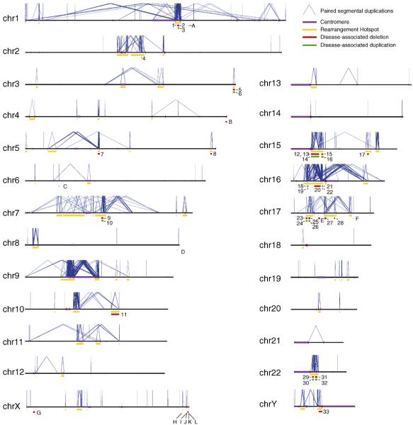
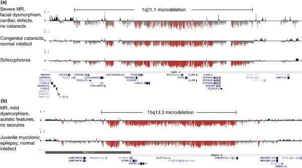
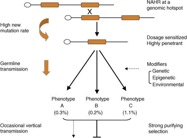
The human genome is enriched in interspersed segmental duplications that sensitize approximately 10% of our genome to recurrent microdeletions and microduplications as a result of unequal crossing over. We review the recent discovery of recurrent rearrangements within these genomic hotspots and their association with both syndromic and nonsyndromic diseases. Studies of common complex genetic disease show that a subset of these recurrent events plays an important role in autism, schizophrenia, and epilepsy. The genomic hotspot model may provide a powerful approach for understanding the role of rare variants in common disease.


Figures

Figure 1
Figure 1
Rearrangement hotspots and their associated disease phenotypes. Each chromosome is depicted as a horizontal line with intrachromosomal segmental duplications connected by blue lines. Gold bars represent “rearrangement hotspots” defined as unique regions (50 kb–10 Mb) flanked by intrachromosomal segmental duplications >10 kb with >95% sequence identity. Disease-associated deletions and duplications are represented by red or green bars, respectively. Numbers represent rearrangements within hotspot regions as defined above: (1) TAR syndrome, (2) 1q21.1 deletion and (3) reciprocal duplication, (4) Juvenile Nephronophthisis, (5) 3q29 deletion and (6) reciprocal duplication, (7) Spinal Muscular Atrophy, (8) Sotos syndrome, (9) Williams syndrome and (10) reciprocal duplication, (11) 10q22–q23 microdeletion, (12) Prader-Willi syndrome, (13) Angelman syndrome, (14) duplication 15q11, (15) 15q13.3 microdeletion and (16) reciprocal duplication, (17) 15q24 microdeletion, (18) 16p13.11 deletion and (19) reciprocal duplication, (20) 16p11.2–p12.2 deletion and (21) 16p11.2 deletion and (22) reciprocal duplication, (23) HNPP, (24) CMT1A, (25) Smith-Magenis syndrome, (26) Potocki-Lupski syndrome, (27) renal cysts and diabetes (RCAD) syndrome, (28) 17q21.31 microdeletion syndrome, (29) velocardiofacial/DiGeorge/deletion 22q11 syndrome and (30) reciprocal duplication, (31) distal 22q11.2 deletion and (32) reciprocal duplication, and (33) azoospermia. Letters represent NAHR-mediated deletions that occur outside of hotspots (as defined above): (A) Gaucher disease, (B) Fascioscapulohumeral dystrophy, (C) congenital adrenal hyperplasia, (D) glucocorticoid-remediable aldosteronism, (E) Neurfibromatosis type I microdeletion syndrome, (F) pituitary dwarfism, (G) X-linked ichthyosis, (H) Hunter syndrome, (I) red-green color-blindness, (J) Emery-Dreifuss muscular dystrophy, (K) incontinentia pigmenti and (L) hemophilia A.
Figure 2
Figure 2
Identical deletions are associated with diverse phenotypic outcomes. Oligonucleotide array CGH results are depicted for (A) chromosome 1q21.1 (hg18, chr1: 143,000,000–145,500,000) and (B) chromosome 15q13.3 (hg18, chr15: 28,000,000–31,000,000). X-axis, genomic location; y-axis, log2 ratio of fluorescence intensity; red and green bars represent log2 ratio less than or greater than 1.5 standard deviations from the mean, respectively. Clinical features for the individuals shown are listed to the left of the array CGH results for that individual. Segmental duplication blocks are depicted by orange/yellow/gray bars at the top (90–94.9%, 95–99% and >99% sequence identity respectively); genes are shown below each figure.
Figure 3
Figure 3
Model for recurrent rearrangements in common disease. Orange blocks represent segmental duplications flanking a unique stretch of sequence. Mis-alignment at meiosis results in non-allelic homologous recombination (NAHR) resulting in deletion (shown) or duplication (not shown). See text for additional details.

References

    1. Butler MG, Meaney FJ, Palmer CG. Clinical and cytogenetic survey of 39 individuals with Prader-Labhart-Willi syndrome. Am J Med Genet. 1986;23:793–809. - PMC - PubMed
    1. Smith AC, McGavran L, Robinson J, Waldstein G, Macfarlane J, Zonona J, Reiss J, Lahr M, Allen L, Magenis E. Interstitial deletion of (17)(p11.2p11.2) in nine patients. Am J Med Genet. 1986;24:393–414. - PubMed
    1. Lupski JR. Genomic disorders: structural features of the genome can lead to DNA rearrangements and human disease traits. Trends Genet. 1998;14:417–422. - PubMed
    2. **Key paper defining “genomic disorders” as conditions that result from DNA rearrangements due to regional genomic architecture.

    1. Lupski JR, de Oca-Luna RM, Slaugenhaupt S, Pentao L, Guzzetta V, Trask BJ, Saucedo-Cardenas O, Barker DF, Killian JM, Garcia CA, et al. DNA duplication associated with Charcot-Marie-Tooth disease type 1A. Cell. 1991;66:219–232. - PubMed
    2. *CMT1A (duplication of 17p11) and HNPP (deletion of 17p11; ref. 5) were the first two genomic disorders described to result from reciprocal rearrangements mediated by segmental duplications.

    1. Chance PF, Alderson MK, Leppig KA, Lensch MW, Matsunami N, Smith B, Swanson PD, Odelberg SJ, Disteche CM, Bird TD. DNA deletion associated with hereditary neuropathy with liability to pressure palsies. Cell. 1993;72:143–151. - PubMed
    2. *CMT1A (duplication of 17p11; ref. 4) and HNPP (deletion of 17p11) were the first two genomic disorders described to result from reciprocal rearrangements mediated by segmental duplications.

Publication types