. 2009 Aug;39(10):1063-70.
doi: 10.1016/j.ijpara.2009.05.004. Epub 2009 May 25.

Conservation of proteins involved in oocyst wall formation in Eimeria maxima, Eimeria tenella and Eimeria acervulina

Affiliations

Conservation of proteins involved in oocyst wall formation in Eimeria maxima, Eimeria tenella and Eimeria acervulina

Sabina I Belli et al. Int J Parasitol. 2009 Aug.

Abstract

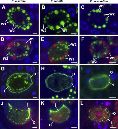
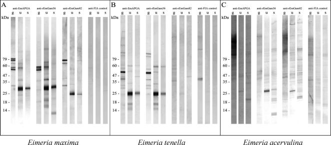
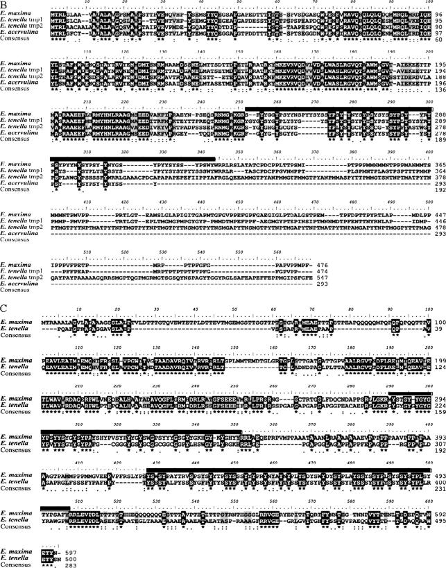
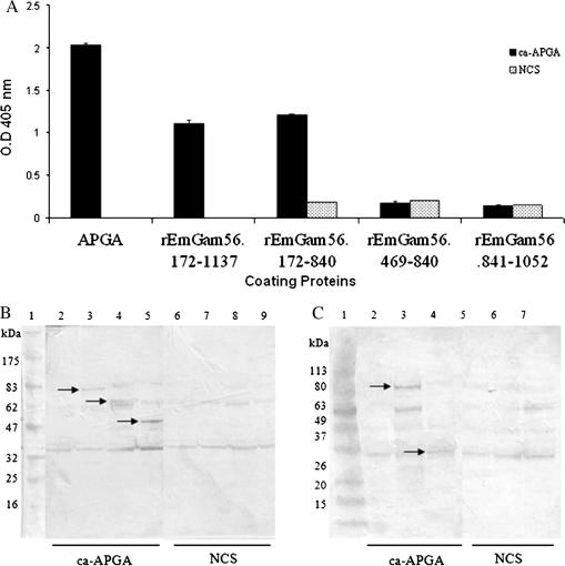
Vaccination with proteins from gametocytes of Eimeria maxima protects chickens, via transfer of maternal antibodies, against infection with several species of Eimeria. Antibodies to E. maxima gametocyte proteins recognise proteins in the wall forming bodies of macrogametocytes and oocyst walls of E. maxima, Eimeria tenella and Eimeria acervulina. Homologous genes for two major gametocyte proteins - GAM56 and GAM82 - were found in E. maxima, E. tenella and E. acervulina. Alignment of the predicted protein sequences of these genes reveals that, as well as sharing regions of tyrosine richness, strong homology exists in their amino-terminal regions, where protective antibodies bind. This study confirms the conservation of the roles of GAM56 and GAM82 in oocyst wall formation and shows that antibodies to gametocyte antigens of E. maxima cross-react with homologous proteins in other species, helping to explain cross-species maternal immunity.


Figures

Fig. 1
Fig. 1
Details of mature macrogametes (A–F) or developing oocysts (G–L) immuno-stained with anti-EmAPGA (antibody to Eimeria maxima Affinity Purified Gametocyte Antigens) (A–C, G–I), visualised with FITC (green) and counter-stained with DAPI (blue) or anti-rEmGAM56 (antibody to recombinant E. maxima 56 kDa gametocyte protein) (D, E, J, K) or anti-rEmGAM82 (antibody to recombinant E. maxima 82 kDa gametocyte protein) (F, L) visualised with FITC and counter-stained with DAPI (blue) and Evans blue (red). The first column (A, D, G, J) shows examples of E. maxima, the second column (B, E, H, K) is Eimeria tenella and the third column (C, F, I, L) is Eimeria acervulina. Bars represent 5 μm. The anti-EmAPGA labels the WFB1 (Wall Forming Body Type 1) (W1) and WFB2 (W2) in all three species (A–C) while anti-rEmGAM56 labels the WFB2 (W2) but not the WFB1 (W1) in E. maxima (D) and E. tenella (E) and anti-rEmGAM82 shows a similar staining pattern in E. acervulina (F). In the oocysts, anti-EmAPGA labels the outer layer (O) and the inner layer (I) of the oocyst wall in all three species (G–I). In contrast, anti-rEmGAM56 in E. maxima (J) and E. tenella (K) and anti-rEmGAM82 in E. acervulina (L) stain the inner layer (I) but not the outer layer of the oocyst wall (© David J.P. Ferhuson).
Fig. 2
Fig. 2
Cross-reactivity of antibodies to Eimeria maxima gametocyte antigens with E.maxima (A) Eimeria tenella (B) and Eimeria acervulina (C). Gametocyte (g), unsporulated oocysts (u) and sporulated oocysts (s) were separated by SDS–PAGE, transferred to Immobilon-P membrane and probed with antisera to E. maxima gametocyte proteins (anti-EmAPGA [antibody to Eimeria maxima Affinity Purified Gametocyte Antigens] anti-rEmGAM56 [antibody to recombinant E. maxima 56 kDa gametocyte protein], anti-rEmGAM82 [antibody to recombinant E. maxima 82 kDa gametocyte protein]) or antisera from animals immunised with Freund’s Incomplete Adjuvant (FIA) plus saline. Bands were visualised with SIGMAFAST™ BCIP/NBT after incubation with secondary antibody. Parasite loadings per well were as follows: E. maxima – 2 × 103 gametocytes, 3 × 103 unsporulated oocysts, 2.5 × 104 sporulated oocysts; E. tenella – 2 × 103 gametocytes, 2 × 103 unsporulated oocysts, 2 × 104 sporulated oocysts; E. acervulina – 7.5 × 103 of gametocytes, 6 × 104 unsporulated oocysts, 2.4 × 105 sporulated oocysts.
Fig. 3
Fig. 3
Stage-specific gene expression of the gam56 gene in Eimeria tenella (A) and multiple protein sequence alignment of GAM56 (56 kDa gametocyte protein) (B) and GAM82 (C) from Eimeria maxima, E. tenella and Eimeria acervulina. Total RNA was extracted from E. tenella life cycle stages, cDNA synthesised and parasite genes amplified by PCR (A). Purified merozoites (1 × 107) and gametocytes (1 × 106) were resuspended in 1 ml TRIzol® Reagent and homogenised by pipetting. Unsporulated oocysts (2 × 105) and sporulated oocysts (5 × 105) were resuspended in 1 ml TRIzol® Reagent and 1 vol. of glass beads was added and the sample was vortexed for 1 min intervals until disruption of oocysts was confirmed by bright field microscopy. All TRIzol® treated samples were left at room temperature for 10 min and total RNA isolated by chloroform extraction and isopropanol precipitation. RNA was quantified using a NanoDrop ND-1000 Spectrophotometer and cDNA was synthesised using SuperScript III Reverse Transcriptase (Invitrogen) according to the manufacturer’s instructions. Parasite cDNA samples were standardised by quantification of E. tenella β-actin PCR product. Forward primer E0043 (5′ ggaattcgttggccgcccaagaatcc 3′) and reverse primer E0044 (5′ gctctagattagctcggcccagactcatc 3′) were used to generate a 1020 bp β-actin cDNA PCR product. Forward primer E0030 (5′ catatggtggagaacacggtgcac 3′) and reverse primer E0031 (5′ ctcgagttagtaccagctggaggagta 3′) were designed to amplify a 906 bp cDNA product of etgam56 tmp 1. Forward primer E0035 (5′ catatggtagaagtgccaatggac 3′) and reverse primer E0036 (5′ cacgtgttagtagaagctggagtggct 3′) were designed to amplify an 804 bp cDNA product of etgam56 tmp 2. Each PCR reaction contained 50 ng of parasite stage-specific cDNA, 0.2 μM forward primer, 0.2 μM reverse primer, 1 × AccuPrime™ reaction mix, and AccuPrimePfx DNA polymerase (Invitrogen). The PCR reaction was carried out as follows: initial denaturation 95 °C for 3 min; 95 °C for 30 s; 61 °C for 1 min; 68 °C for 1.5 min, for 25 cycles with a final extension at 68 °C for 10 min. Merozoite (M), gametocyte (G), unsporulated oocyst (UO), sporulated oocyst (SO), genomic DNA control (gD) and a negative control sample with no template (C) were electrophoresed on a 1% agarose gel alongside a 100 bp ladder (Invitrogen) and visualised using Gel Red™ (Biotium). The β-actin gene product (∼1000 bp) was amplified from cDNA from all E. tenella life cycle stages. The etgam56 tmp 1 gene product (∼900 bp) and etgam56 tmp 2 gene product (∼800 bp) were amplified from cDNA of E. tenella gametocytes and the genomic DNA control only. The amino acid sequences of GAM56 and GAM82 were aligned using the program Clustal X and BLOSUM (BLOcks of amino acid Substitution Matrix) (Chenna et al., 2003) (B and C). Tyrosine rich regions (highlighted with a black line above the alignment) split the protein sequences into the N- and C-terminal ends. Symbols on the lowermost Clustal consensus lines represent amino acid positions: ‘∗’ fully conserved, ‘:’ one of the strong groups of amino acids is conserved, and ‘.’ one of the weak amino acid groups is conserved.
Fig. 3
Fig. 3
Stage-specific gene expression of the gam56 gene in Eimeria tenella (A) and multiple protein sequence alignment of GAM56 (56 kDa gametocyte protein) (B) and GAM82 (C) from Eimeria maxima, E. tenella and Eimeria acervulina. Total RNA was extracted from E. tenella life cycle stages, cDNA synthesised and parasite genes amplified by PCR (A). Purified merozoites (1 × 107) and gametocytes (1 × 106) were resuspended in 1 ml TRIzol® Reagent and homogenised by pipetting. Unsporulated oocysts (2 × 105) and sporulated oocysts (5 × 105) were resuspended in 1 ml TRIzol® Reagent and 1 vol. of glass beads was added and the sample was vortexed for 1 min intervals until disruption of oocysts was confirmed by bright field microscopy. All TRIzol® treated samples were left at room temperature for 10 min and total RNA isolated by chloroform extraction and isopropanol precipitation. RNA was quantified using a NanoDrop ND-1000 Spectrophotometer and cDNA was synthesised using SuperScript III Reverse Transcriptase (Invitrogen) according to the manufacturer’s instructions. Parasite cDNA samples were standardised by quantification of E. tenella β-actin PCR product. Forward primer E0043 (5′ ggaattcgttggccgcccaagaatcc 3′) and reverse primer E0044 (5′ gctctagattagctcggcccagactcatc 3′) were used to generate a 1020 bp β-actin cDNA PCR product. Forward primer E0030 (5′ catatggtggagaacacggtgcac 3′) and reverse primer E0031 (5′ ctcgagttagtaccagctggaggagta 3′) were designed to amplify a 906 bp cDNA product of etgam56 tmp 1. Forward primer E0035 (5′ catatggtagaagtgccaatggac 3′) and reverse primer E0036 (5′ cacgtgttagtagaagctggagtggct 3′) were designed to amplify an 804 bp cDNA product of etgam56 tmp 2. Each PCR reaction contained 50 ng of parasite stage-specific cDNA, 0.2 μM forward primer, 0.2 μM reverse primer, 1 × AccuPrime™ reaction mix, and AccuPrimePfx DNA polymerase (Invitrogen). The PCR reaction was carried out as follows: initial denaturation 95 °C for 3 min; 95 °C for 30 s; 61 °C for 1 min; 68 °C for 1.5 min, for 25 cycles with a final extension at 68 °C for 10 min. Merozoite (M), gametocyte (G), unsporulated oocyst (UO), sporulated oocyst (SO), genomic DNA control (gD) and a negative control sample with no template (C) were electrophoresed on a 1% agarose gel alongside a 100 bp ladder (Invitrogen) and visualised using Gel Red™ (Biotium). The β-actin gene product (∼1000 bp) was amplified from cDNA from all E. tenella life cycle stages. The etgam56 tmp 1 gene product (∼900 bp) and etgam56 tmp 2 gene product (∼800 bp) were amplified from cDNA of E. tenella gametocytes and the genomic DNA control only. The amino acid sequences of GAM56 and GAM82 were aligned using the program Clustal X and BLOSUM (BLOcks of amino acid Substitution Matrix) (Chenna et al., 2003) (B and C). Tyrosine rich regions (highlighted with a black line above the alignment) split the protein sequences into the N- and C-terminal ends. Symbols on the lowermost Clustal consensus lines represent amino acid positions: ‘∗’ fully conserved, ‘:’ one of the strong groups of amino acids is conserved, and ‘.’ one of the weak amino acid groups is conserved.
Fig. 4
Fig. 4
Analyses of the antigenicity of truncated recombinant proteins of Eimeria maxima GAM56 (EmGAM56; E. maxima 56 kDa gametocyte protein) (A) and EmGAM82 (E. maxima 82 kDa gametocyte protein) (B, C). The recombinant versions of emgam56 (designated remgam56.172-1137 here) and emgam82 (designated remgam82.168-1887 here) described by Belli et al. (2002b, 2003b, 2004) served as parent constructs for DNA amplification of various regions by PCR. DNA fragments were amplified from the parent construct using specific primer sets and designated according to the first and last base pairs included in their sequences: remgam56.172-840 (forward primer = 5′ cgcggatccgaccactcctgtggagaatcaggt 3′, reverse primer = 5′ cgcgaattcgatcatgtccatcatctcggtaac 3′); remgam56.469-840 (forward primer = 5′ cgcggatccgtccaacagaatgaatgcagcaat 3′, reverse primer = 5′ ggcgaattcgctcctgccctttctgcccatatt 3′); remgam56.172-840 (forward primer = 5′ cgcggatccgaccactcctgtggagaatcaggt 3′, reverse primer = 5′ ggcgaattcgctcctgccctttctgcccatatt 3′); remgam56.841-1052 (forward primer = 5′ cgcggatccgttctactcctgcggctatcccag 3′, reverse primer = 5′ cgcgaattcgctggggtagctgctataactgta 3′); remgam82.168-1620 (forward primer = 5′ cgcggatcctactgtattggacacaacgactggc 3′, reverse primer = 5′ cgcgaattcatcagggacctctagtctttctataaaagg 3′); remgam82.168-1169 (forward primer = 5′ cgcggatcctactgtattggacacaacgactggc 3′, reverse primer = 5′ cgcgaattcgcataacaggtcttggttcctgctc 3′); and remgam82.168-824 1169 (forward primer = 5′ cgcggatcctactgtattggacacaacgactggc 3′, reverse primer = 5′ ggcgaattcgcacgaagacgatcatgcatgcga 3′). Purified recombinant proteins of EmGAM56 were coated onto ELISA plates (100 ng/well) and exposed to 100 μl of chicken anti-EmAPGA (E. maxima Affinity Purified Gametocyte Antigens) serum (ca-APGA; 1:100 dilution) or negative control chicken serum (NCS) and assayed using methods described previously (Belli et al., 2004) (B). Results are the means of duplicate assays. For EmGAM82, induced recombinant bacterial lysates were separated by SDS–PAGE and proteins transferred to polyvinylidene fluoride membrane and probed with chicken anti-EmAPGA serum (ca-APGA), and negative control chicken serum (NCS) using protocols described previously (Belli et al., 2004) (B and C). Lanes for (B) are: (1) protein molecular weight markers; (2 and 6) bacterial lysate of induced pTrcHisB vector; (3 and 7) bacterial lysate of induced remgam82.168-1887 in pTrcHisB; (4 and 8) bacterial lysate of induced remgam82.168-1620 in pTrcHisB; (5 and 9) bacterial lysate of induced remgam82.168-1169 in pTrcHisB. Lanes for (C) are: (1) benchmark prestained protein molecular weight markers; (2 and 5) bacterial lysate of induced pTrcHisB vector; (3 and 6) bacterial lysate of induced remgam82.168-1887 in pTrcHisB; (4 and 7) bacterial lysate of induced remgam82.168-824 in pTrcHisB.

References

    1. Belli S.I., Lee M., Thebo P., Wallach M.G., Schwartsburd B., Smith N.C. Biochemical characterisation of the 56 and 82 kDa immunodominant gametocyte antigens from Eimeria maxima. Int. J. Parasitol. 2002;32:805–816. - PubMed
    1. Belli S.I., Witcombe D., Wallach M.G., Smith N.C. Functional genomics of gam56: characterisation of the role of a 56 kDa sexual stage antigen in oocyst wall formation in Eimeria maxima. Int. J. Parasitol. 2002;32:1727–1737. - PubMed
    1. Belli S.I., Wallach M.G., Luxford C., Davies M.J., Smith N.C. Roles of tyrosine-rich precursor glycoproteins and dityrosine- and 3,4-dihydroxyphenylalanine-mediated protein cross-linking in development of the oocyst wall in the coccidian parasite Eimeria maxima. Eukaryot. Cell. 2003;2:456–464. - PMC - PubMed
    1. Belli S.I., Wallach M.G., Smith N.C. Cloning and characterization of the 82 kDa tyrosine-rich sexual stage glycoprotein, GAM82, and its role in oocyst wall formation in the apicomplexan parasite, Eimeria maxima. Gene. 2003;307:201–212. - PubMed
    1. Belli S.I., Mai K., Skene C.D., Gleeson M.T., Witcombe D.M., Katrib M., Finger A., Wallach M.G., Smith N.C. Characterisation of the antigenic and immunogenic properties of bacterially expressed, sexual stage antigens of the coccidian parasite, Eimeria maxima. Vaccine. 2004;22:4316–4325. - PubMed

Publication types

LinkOut - more resources