

Regulation of superoxide dismutase genes: implications in disease
- PMID: 19477268
- PMCID: PMC2731574
- DOI: 10.1016/j.freeradbiomed.2009.05.018
Regulation of superoxide dismutase genes: implications in disease
Abstract
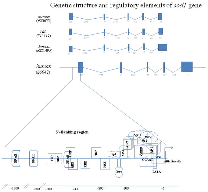
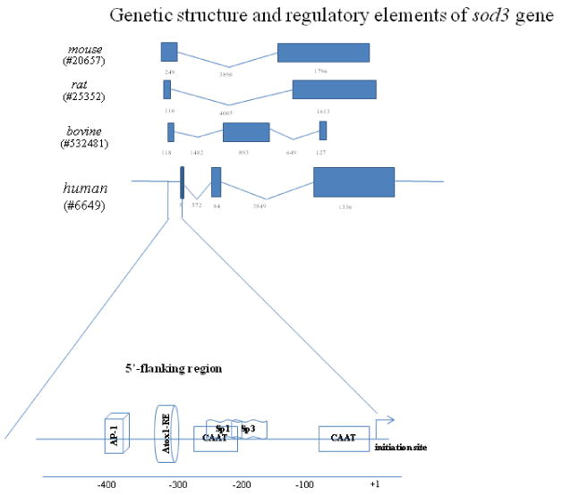
Numerous short-lived and highly reactive oxygen species (ROS) such as superoxide (O2(.-)), hydroxyl radical, and hydrogen peroxide are continuously generated in vivo. Depending upon concentration, location, and intracellular conditions, ROS can cause toxicity or act as signaling molecules. The cellular levels of ROS are controlled by antioxidant enzymes and small-molecule antioxidants. As major antioxidant enzymes, superoxide dismutases (SODs), including copper-zinc superoxide dismutase (Cu/ZnSOD), manganese superoxide dismutase, and extracellular superoxide dismutase, play a crucial role in scavenging O2(.-). This review focuses on the regulation of the sod genes coding for these enzymes, with an emphasis on the human genes. Current knowledge about sod structure and regulation is summarized and depicted as diagrams. Studies to date on genes coding for Cu/ZnSOD (sod1) are mostly focused on alterations in the coding region and their associations with amyotrophic lateral sclerosis. Evaluation of nucleotide sequences reveals that regulatory elements of the sod2 gene reside in both the noncoding and the coding region. Changes associated with sod2 lead to alterations in expression levels as well as protein function. We also discuss the structural basis for the changes in SOD expression associated with pathological conditions and where more work is needed to establish the relationship between SODs and diseases.
Figures

References
-
- Evans MD, Dizdaroglu M, Cooke MS. Oxidative DNA damage and disease: induction, repair and significance. Mutat Res. 2004;567:1–61. - PubMed
-
- Xie L, Zhu X, Hu Y, Li T, Gao Y, Shi Y, Tang S. Mitochondrial DNA oxidative damage triggering mitochondrial dysfunction and apoptosis in high glucose-induced HRECs. Invest Ophthalmol Vis Sci. 2008;49:4203–4209. - PubMed
-
- Sun Y, Oberley LW. Redox regulation of transcriptional activators. Free Radic Biol Med. 1996;21:335–348. - PubMed
-
- Cai J, Yang J, Jones DP. Mitochondrial control of apoptosis: the role of cytochrome c. Biochim Biophys Acta. 1998;1366:139–149. - PubMed
Publication types
MeSH terms
Substances
Grants and funding
LinkOut - more resources
Full Text Sources
Medical
Miscellaneous
