. 2009 Jun 2;15(6):539-50.
doi: 10.1016/j.ccr.2009.03.027.

Expression of autotaxin and lysophosphatidic acid receptors increases mammary tumorigenesis, invasion, and metastases

Affiliations

Expression of autotaxin and lysophosphatidic acid receptors increases mammary tumorigenesis, invasion, and metastases

Shuying Liu et al. Cancer Cell. .

Erratum in

  • Cancer Cell. 2009 Aug 4;16(2):172

Abstract

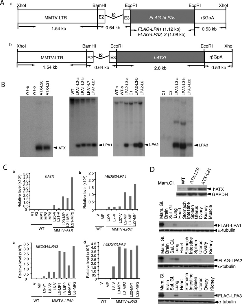
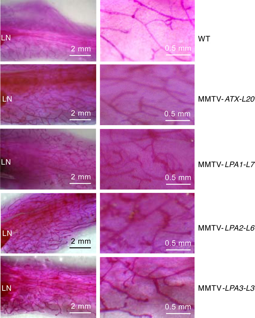
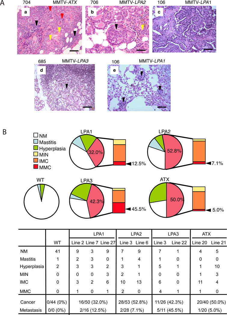
Lysophosphatidic acid (LPA) acts through high-affinity G protein-coupled receptors to mediate a plethora of physiological and pathological activities associated with tumorigenesis. LPA receptors and autotaxin (ATX/LysoPLD), the primary enzyme producing LPA, are aberrantly expressed in multiple cancer lineages. However, the role of ATX and LPA receptors in the initiation and progression of breast cancer has not been evaluated. We demonstrate that expression of ATX or each edg family LPA receptor in mammary epithelium of transgenic mice is sufficient to induce a high frequency of late-onset, estrogen receptor (ER)-positive, invasive, and metastatic mammary cancer. Thus, ATX and LPA receptors can contribute to the initiation and progression of breast cancer.


Figures

Figure 1
Figure 1. Generation and characterization of transgenic mice
(A) The MMTV-LPA1, 2, 3 (a) or MMTV-ATX (b) transgenic constructs contain the 1.54 kb MMTV-LTR and the full-length cDNA of FLAG-tagged human LPA receptors or full-length human ATX linked to the KCR fragment containing the exon II (E2), intron II (I2), exon III (E3) and polyadenylation signal (rβGpA) derived from rabbit β-globin. (B) Southern analysis of BamHI/XhoI digested genomic DNA (15 µg) from potential founder mice hybridized with full-length human LPA1, 2, 3 cDNA probes or StuI/EcoRI fragment of hATX, genomic DNA from wild type FVB/N (WT), or other strain of edg family as control (C1, LPA1; C2, LPA2) for MMTV-LPAs. (C). ATX or LPA receptor transgene RNA expression in mammary glands of virgin (V) and multiparous mice (MP, at day 18 of the 2nd pregnancy) from different lines (L) were analyzed by Real-time quantitative PCR. D. ATX or LPA receptor protein in transgenic mice, mammary gland (Mam. Gl), Salivary gland (Sal. Gl).
Figure 2
Figure 2. Effect of transgene expression in virgin murine mammary glands
Whole mount analyses of the fourth inguinal mammary glands of 8-week-old virgin mice. Results are representative images from 6 mice of each group. The right column is magnification of the left column. LN indicates lymph node.
Figure 3
Figure 3. Mammary cancer incidence in female MMTV-ATX, or MMTV-LPA1, 2, 3 transgenic mice
Female transgenic mice were bred 3 times to induce the MMTV-LTR promoter and monitored for tumor incidence over 24 months with WT females including FVB/N and nontransgenic littermates as controls. (A) Mammary cancers, large (1.5 × 2.0 × 1.6 cm, black arrowhead) and small (0.5 × 0.5 × 0.5 cm, red arrowhead), developed in MMTV-LPA2. (B) Kaplan-Meyer analysis of mammary cancers. (C) Mammary cancer incidence in each line.
Figure 4
Figure 4. Spectrum of mammary gland lesions
(A) Histopathology of mammary gland lesions in MMTV-LPAs or MMTV-ATX transgenic mice. (a) Chronic mastitis with white blood cell infiltration (black arrowhead) & squamous metaplasia (yellow arrowhead) of hyperplastic ducts (red arrowhead). (b) Mammary intraepithelial neoplasia (MIN) (black arrowhead) with hyperplastic ducts (yellow arrowhead) and chronic mastitis. (c) Invasive mammary adenocarcinoma. (d) Lymph node metastatic carcinoma (black arrowhead). (e) Lung metastatic carcinoma (black arrowhead) with pneumonia (scale 50 µm). (B) Representative mammary gland lesion histopathology. NM, normal mammary gland; Mastitis, chronic mastitis with squamous metaplasia; Hyperplasia, hyperplasia (with chronic mastitis); MIN, mammary intraepithelial neoplasia (with chronic mastitis and hyperplasia); IMC, invasive mammary carcinoma (with mastitis, hyperplasia or MIN); MMC, metastatic mammary carcinoma.
Figure 5
Figure 5. ER expression in mammary glands and mammary carcinomas from MMTV–LPAs or MMTV-ATX
Paraffin sections of mammary carcinomas or mammary glands from adult transgenic mice (20–22 month-old multiparous) were assessed for ER by immunohistochemical staining, with normal mammary glands from WT multiparous mice as control (including FVB/N and nontransgenic littermates). (A) ER expression in mammary glands of wild type FVB (a), transgenic mice (b), or mammary ductal hyperplasia (c–e), (scale 50 µm). (B) ER expression in tumors. (a) Mammary adenocarcinoma from MMTV-ATX; (b) Mammary adenocarcinoma from MMTV-LPA1; (c) Mammary lymph node metastatic adenocarcinoma from MMTV-ATX; (d) Lung metastatic adenocarcinoma from MMTV-LPA3 (scale 100 µm).
Figure 6
Figure 6. MIP-3α and VEGF production in MMTV-ATX or MMTV-LPA1, 2, 3 mice
Serum MIP-3α and VEGF in transgenic mice with and without mammary carcinoma (MC) were quantified using MIP-3α and VEGF ELISA Kit. MIP-3α and VEGF concentrations were calculated by comparing the absorbance of samples to standard curves. (A) MIP-3α; (B) VEGF.
Figure 7
Figure 7. Effects of transgene expression on cellular signaling pathways
(A) Variation in expression or phosphorylation of 50 proteins in 152 experimental samples from 145 mice. Data are presented in a matrix format: each row represents an experimental sample, and each column an antibody target. In each sample, the ratio of the abundance of the molecule to its median abundance across all tissue samples is represented by the color of the corresponding cell in the matrix (see scale, for expression levels). Dendrogram on left shows similarities in the expression patterns between experimental samples. (B) Phosphorylation of p38-MAPK, ERK/MAPK, ATF-2, NF-κB, pPKCα, AKT, GSK3α/β and S6 in mammary carcinomas (MC) from MMTV-ATX and MMTV-LPA1 2, 3 mice was analyzed by Western blot. (C) Immunohistochemistry was performed on paraffin-embedded mammary carcinoma sections. Specimens were blocked and incubated with rabbit polyclonal antibody to β-catenin (detailed in Experimental procedures). β-catenin expression in normal mammary gland of wild type female mice (a); mammary adenocarcinoma from MMTV-LPA2 (b); mammary lymph node metastatic adenocarcinoma from MMTV-LPA2 and MMTV-ATX (c & d) (scale 50 µm).

Comment in

References

    1. Aylon Y, Oren M. Living with p53, Dying of p53. Cell. 2007;130:597–600. - PubMed
    1. Baker DL, Fujiwara Y, Pigg KR, Tsukahara R, Kobayashi S, Murofushi H, Uchiyama A, Murakami-Koh E, Bandle RW, et al. Carba analogs of cyclin phophatidic acid are selective inhibitors of autotaxin and cancer cell invasion and metastasis. J Biol Chem. 2006;281:22786–22793. - PMC - PubMed
    1. Balkwill F, Charles KA, Mantovani A. Smoldering and polarized inflammation in the initiation and promotion of malignant disease. Cancer Cell. 2005;7:211–217. - PubMed
    1. Benito M, Parker J, Du Q, Wu J, Xiang D, Perou CM, Marron JS. Adjustment of systematic microarray data biases. Bioinformatics. 2004;20:105–114. - PubMed
    1. Benjamini Y, Hochberg Y. Controlling the false discovery rate: a practical and powerful approach to multiple testing. J R Statist Soc B. 1995;57:289–300.

Publication types

MeSH terms

Substances

Associated data