

Review
doi: 10.1016/j.sbi.2009.04.006.
Epub 2009 May 26.
Structural and functional modules in RNA interference
Affiliations
- PMID: 19477631
- PMCID: PMC2721689
- DOI: 10.1016/j.sbi.2009.04.006
Item in Clipboard
Review
Structural and functional modules in RNA interference
Curr Opin Struct Biol.
2009 Jun.
Abstract
RNA interference (RNAi) uses small RNA molecules to regulate transcriptional and post-transcriptional gene expression. In recent years, a number of structural studies provided insights into the molecular architecture and mechanism of functional modules of RNAi. Mechanisms of nucleic acid recognition and cleavage have been revealed by structural studies of proteins and their nucleic acid complexes involved in RNA biogenesis, for example, Argonaute, PIWI, RNase III, Dicer, Drosha, and DGCR8. While quite a few questions remain, an excellent structural and mechanistic overview of RNAi processes has already emerged. In this review, we examine functional modules and their assemblies in RNAi processes.
Figures
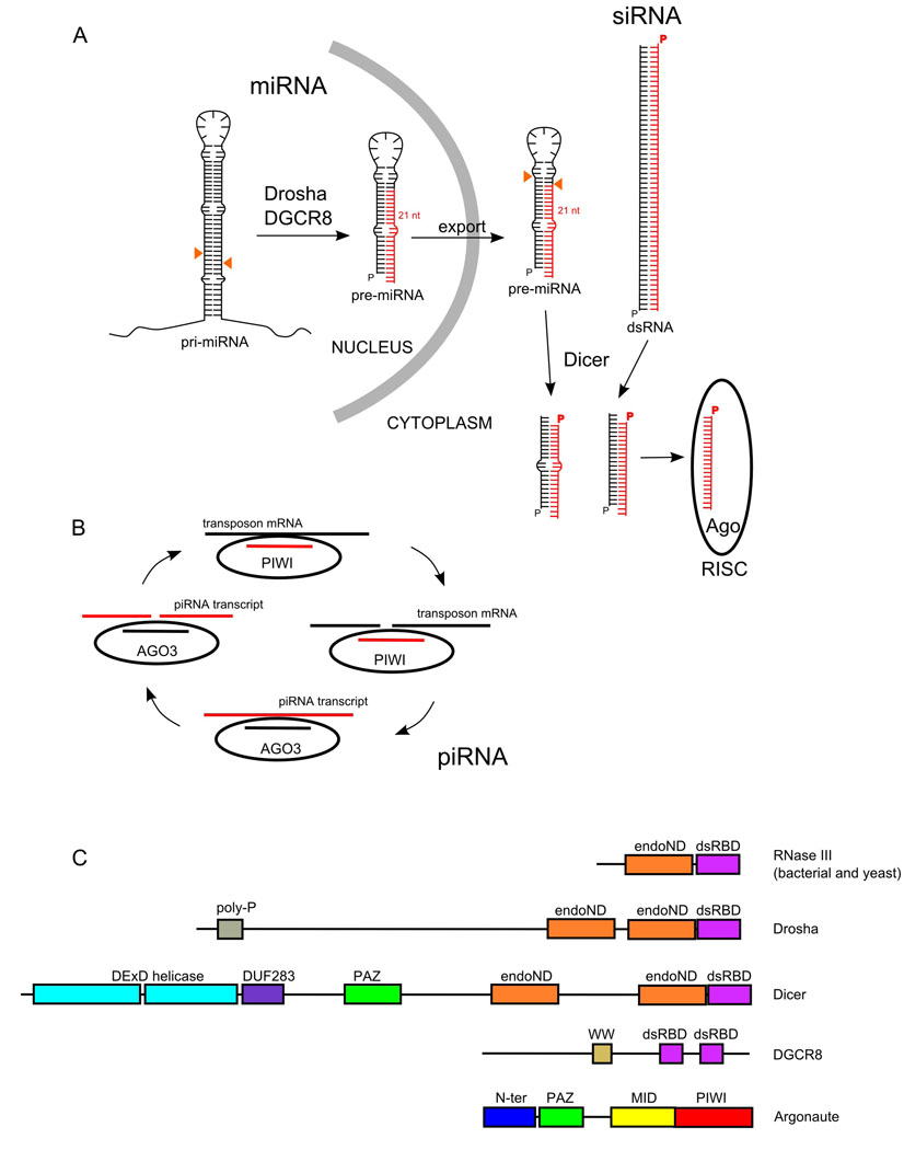
(A) Processing of RNAs in miRNA and siRNA. Pri-miRNA transcripts are processed by Drosha/DGCR8 in the nucleus and Dicer in the cytoplasm. The cleavage sites are indicated by orange triangles. siRNAs start as dsRNA and are processed by Dicer. In both cases the end product 20–30 nt RNA is incorporated into the RISC complex. (B) Model of piRNA pathway. Transposon mRNA is cleaved by PIWI using a piRNA guide strand. A fragment of cleaved transposon RNA is incorporated into Ago3 RISC and used to cleave the piRNA cluster transcript. Its fragment is incorporated into PIWI protein to start another round of the cycle. (C) Domain composition of proteins involved in RNA interference.
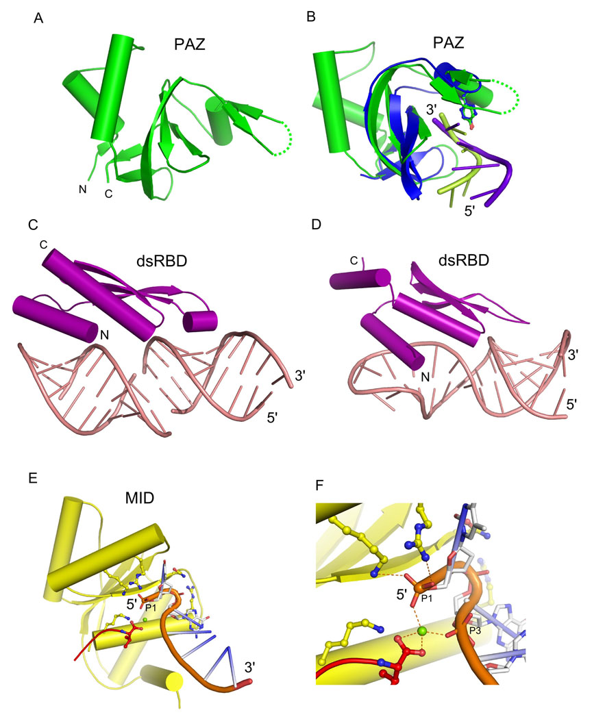
(A) PAZ domain of human Argonaute eIF2c1 (PDB: 1SI3). (B) Comparison of nucleic acid binding by PAZ domains from eIF2c1 (green) and T. thermophilus Ago (PDB: 3DLH) (blue). The nucleic acid from each structure is shown in cartoon representation (light green for eIF2c1 structure and purple blue for T. thermophilus Ago structure). The conserved tyrosine that binds the nucleic acid backbone is shown in ball-and-stick representation. (C) dsRBD (ds-RNA-binding domain) from X. leavis RNA-binding protein A bound to a dsRNA duplex (PDB: 1DI2). (D) dsRBD from Rnt1 bound to a tetraloop-stem RNA (PDB: 1T4L). (E) Mid domain from A. fulgidus PIWI protein bound to ssRNA (PDB: 1YTU). The C-terminal carboxylate is shown in red and the metal ion coordinating the 5’-phosphate of the RNA in green. Conserved residues stabilizing the 5’ end of the RNA are shown in balls-and-sticks. (F) Close-up view of the interactions stabilizing the 5’ end of the RNA.
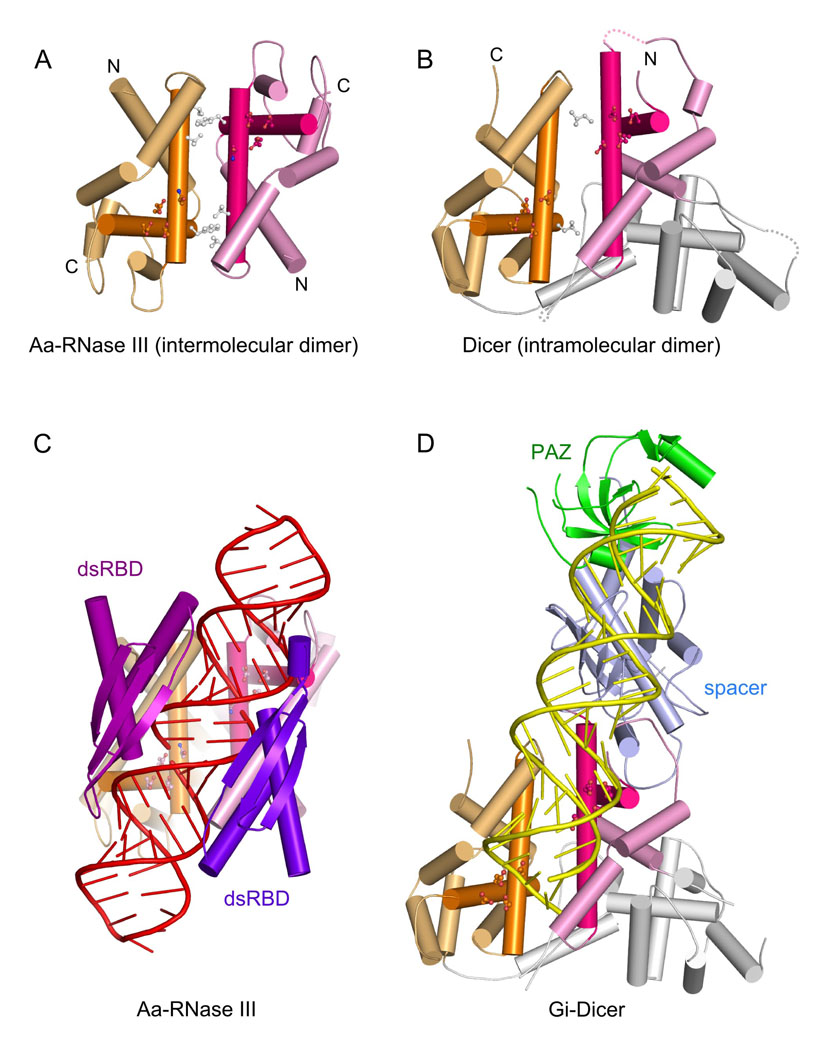
(A) A. aeolicus RNase III – intermolecular homodimer. The two endoNDs from two subunits are shown in orange and pink with the central perpendicular helices shown in darker colors. The residues forming the active site are shown in colored balls-and-sticks and those forming the hydrophobic core of the dimer interface in white balls-and-sticks. (B) G. intestinalis Dicer – the intramolecular dimerization of two endoND domains. The linker domain inserted between them is shown in light grey. (C) The structure of A. aeolicus RNase III in complex with dsRNA (PDB: 2NUF). dsRBDs from each dimer subunit are in purple and blue. The RNA is shown in red. (D) The structure of G. intestinalis Dicer (PDB: 2FFL). The PAZ domain is shown in green and the platform domain in light blue. dsRNA modeled to interact with the endoND and PAZ domains is shown in yellow.
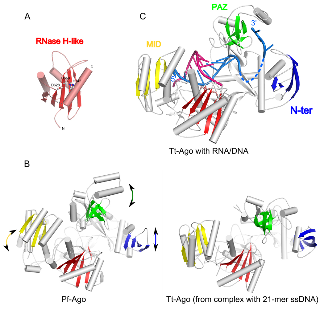
(A) The RNase H-like PIWI domain of P. furiosus Argonaute. The central β-sheet is shown in red and the catalytic residues – two aspartates and a histidine as balls-and-sticks. The site of an ~80 residue insertion into the RNase H fold, which is mainly composed of β-strands, is indicated with a gray dashed line. (B) Comparison of apo P. furiosus Ago structure (left, PDB: 1Z25) and T. thermophilus Ago in complex with 21-nt ss DNA (right, PDB: 3DLH). The protein structures were superimposed using the positions of the active site residues in PIWI (RNase H-like) domain. The central β-sheets of nucleic acid binding domains are shown in color (N-ter - blue, PAZ -green, Mid - yellow, and RNase H-like in red) and the differences in their positions between the two structures are indicated with arrows. (C) Structure of T. thermophilus Ago complexed with an RNA/DNA hybrid (PDB: 3F73). The nucleic acid is shown in cartoon representation (RNA in pink and DNA in light blue). The central β-sheets of each domain are colored as in B. The active site carboxylates are shown in ball-and-sticks.
Similar articles
-
Binding and cleavage specificities of human Argonaute2.J Biol Chem. 2009 Sep 18;284(38):26017-28. doi: 10.1074/jbc.M109.010835. Epub 2009 Jul 22. J Biol Chem. 2009. PMID: 19625255 Free PMC article.
-
Molecular dynamics simulation in RNA interference.Curr Med Chem. 2014 Jun;21(17):1968-75. doi: 10.2174/0929867321666131218100234. Curr Med Chem. 2014. PMID: 24350843 Review.
-
An unusual Dicer-like1 protein fuels the RNA interference pathway in Trypanosoma brucei.RNA. 2006 Dec;12(12):2063-72. doi: 10.1261/rna.246906. Epub 2006 Oct 19. RNA. 2006. PMID: 17053086 Free PMC article.
-
Characterization of the interactions between mammalian PAZ PIWI domain proteins and Dicer.EMBO Rep. 2004 Feb;5(2):189-94. doi: 10.1038/sj.embor.7400070. Epub 2004 Jan 16. EMBO Rep. 2004. PMID: 14749716 Free PMC article.
-
Dicer-independent processing of small RNA duplexes: mechanistic insights and applications.Nucleic Acids Res. 2017 Oct 13;45(18):10369-10379. doi: 10.1093/nar/gkx779. Nucleic Acids Res. 2017. PMID: 28977573 Free PMC article. Review.
Cited by
-
A simple Bayesian estimate of direct RNAi gene regulation events from differential gene expression profiles.BMC Genomics. 2011 May 20;12:250. doi: 10.1186/1471-2164-12-250. BMC Genomics. 2011. PMID: 21599879 Free PMC article.
-
Germ-line deletion in DICER1 revealed by a novel MLPA assay using synthetic oligonucleotides.Eur J Hum Genet. 2014 Apr;22(4):564-7. doi: 10.1038/ejhg.2013.215. Epub 2013 Sep 25. Eur J Hum Genet. 2014. PMID: 24065110 Free PMC article.
-
Novel endoribonucleases as central players in various pathways of eukaryotic RNA metabolism.RNA. 2010 Sep;16(9):1692-724. doi: 10.1261/rna.2237610. Epub 2010 Jul 30. RNA. 2010. PMID: 20675404 Free PMC article. Review.
-
Development of RNA Interference Trigger-Mediated Gene Silencing in Entamoeba invadens.Infect Immun. 2016 Mar 24;84(4):964-975. doi: 10.1128/IAI.01161-15. Print 2016 Apr. Infect Immun. 2016. PMID: 26787723 Free PMC article.
-
Nucleases: diversity of structure, function and mechanism.Q Rev Biophys. 2011 Feb;44(1):1-93. doi: 10.1017/S0033583510000181. Epub 2010 Sep 21. Q Rev Biophys. 2011. PMID: 20854710 Free PMC article. Review.
References
-
- Bushati N, Cohen SM. microRNA functions. Annu Rev Cell Dev Biol. 2007;23:175–205. - PubMed
-
- Filipowicz W, Bhattacharyya SN, sonenberg N. Mechanisms of posttranscriptional regulation by microRNAs: are the answers in sight? Nat Rev Genet. 2008;9:102–114. - PubMed
-
- Rana TM. Illuminating the silence: understanding the structure and function of small RNAs. Nat Rev Mol Cell Biol. 2007;8:23–36. - PubMed
-
- Chapman EJ, Carrington JC. Specialization and evolution of endogenous small RNA pathways. Nat Rev Genet. 2007;8:884–896. - PubMed
-
- Zamore PD, Haley B. the big world of small RNAs. Science. 2005;309:1519–1524. - PubMed
Publication types
MeSH terms
Substances
Grants and funding
LinkOut - more resources
Full Text Sources
Other Literature Sources
