

Synthetic resveratrol aliphatic acid inhibits TLR2-mediated apoptosis and an involvement of Akt/GSK3beta pathway
- PMID: 19477653
- PMCID: PMC2746461
- DOI: 10.1016/j.bmc.2009.05.029
Synthetic resveratrol aliphatic acid inhibits TLR2-mediated apoptosis and an involvement of Akt/GSK3beta pathway
Abstract
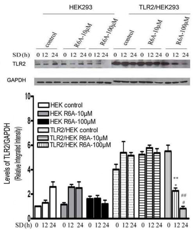
As resveratrol derivatives, resveratrol aliphatic acids were synthesized in our laboratory. Previously, we reported the improved pharmaceutical properties of the compounds compared to resveratrol, including better solubility in water and much tighter binding with human serum albumin. Here, we investigate the role of resveratrol aliphatic acids in Toll-like receptor 2 (TLR2)-mediated apoptosis. We showed that resveratrol aliphatic acid (R6A) significantly inhibits the expression of TLR2. In addition, overexpression of TLR2 in HEK293 cells caused a significant decrease in apoptosis after R6A treatment. Moreover, inhibition of TLR2 by R6A decreases serum deprivation-reduced the levels of phosphorylated Akt and phosphorylated glycogen synthase kinase 3beta (GSK3beta). Our study thus demonstrates that the resveratrol aliphatic acid inhibits cell apoptosis through TLR2 by the involvement of Akt/GSK3beta pathway.
Figures


Similar articles
-
The JAK2-Akt-glycogen synthase kinase-3β signaling pathway is involved in toll-like receptor 2-induced monocyte chemoattractant protein-1 regulation.Mol Med Rep. 2012 Apr;5(4):1063-7. doi: 10.3892/mmr.2012.741. Epub 2012 Jan 3. Mol Med Rep. 2012. PMID: 22218715 Free PMC article.
-
Beta-arrestin 2 modulates resveratrol-induced apoptosis and regulation of Akt/GSK3ß pathways.Biochim Biophys Acta. 2010 Sep;1800(9):912-8. doi: 10.1016/j.bbagen.2010.04.015. Epub 2010 May 7. Biochim Biophys Acta. 2010. PMID: 20457218
-
Resveratrol prevents CA1 neurons against ischemic injury by parallel modulation of both GSK-3β and CREB through PI3-K/Akt pathways.Eur J Neurosci. 2012 Oct;36(7):2899-905. doi: 10.1111/j.1460-9568.2012.08229.x. Epub 2012 Jul 22. Eur J Neurosci. 2012. PMID: 22817531
-
Resveratrol triggers ER stress-mediated apoptosis by disrupting N-linked glycosylation of proteins in ovarian cancer cells.Cancer Lett. 2016 Feb 28;371(2):347-53. doi: 10.1016/j.canlet.2015.11.032. Epub 2015 Dec 15. Cancer Lett. 2016. PMID: 26704305
-
TLR2 ligands induce cardioprotection against ischaemia/reperfusion injury through a PI3K/Akt-dependent mechanism.Cardiovasc Res. 2010 Sep 1;87(4):694-703. doi: 10.1093/cvr/cvq116. Epub 2010 Apr 26. Cardiovasc Res. 2010. PMID: 20421349 Free PMC article.
Cited by
-
Critical role of toll-like receptor 9 in morphine and Mycobacterium tuberculosis-Induced apoptosis in mice.PLoS One. 2010 Feb 19;5(2):e9205. doi: 10.1371/journal.pone.0009205. PLoS One. 2010. PMID: 20174568 Free PMC article.
-
Suppression of inflammatory response by flurbiprofen following focal cerebral ischemia involves the NF-κB signaling pathway.Int J Clin Exp Med. 2014 Sep 15;7(9):3087-95. eCollection 2014. Int J Clin Exp Med. 2014. PMID: 25356186 Free PMC article.
-
Glycogen synthase kinase-3 and p38 MAPK are required for opioid-induced microglia apoptosis.Neuropharmacology. 2010 Nov;59(6):444-51. doi: 10.1016/j.neuropharm.2010.06.006. Epub 2010 Jun 22. Neuropharmacology. 2010. PMID: 20600172 Free PMC article.
-
Hydroxysafflor yellow A protects against cerebral ischemia-reperfusion injury by anti-apoptotic effect through PI3K/Akt/GSK3β pathway in rat.Neurochem Res. 2013 Nov;38(11):2268-75. doi: 10.1007/s11064-013-1135-8. Epub 2013 Aug 29. Neurochem Res. 2013. PMID: 23990223
-
Overexpression of adrenomedullin protects mesenchymal stem cells against hypoxia and serum deprivation‑induced apoptosis via the Akt/GSK3β and Bcl‑2 signaling pathways.Int J Mol Med. 2018 Jun;41(6):3342-3352. doi: 10.3892/ijmm.2018.3533. Epub 2018 Mar 5. Int J Mol Med. 2018. PMID: 29512737 Free PMC article.
References
-
- Pozo-Guisado E, Alvarez-Barrientos A, Mulero-Navarro S, Santiago-Josefat B, Fernandez-Salguero PM. Biochem Pharmacol. 2002;64:1375. - PubMed
-
- Hudson TS, Hartle DK, Hursting SD, Nunez NP, Wang TT, Young HA, Arany P, Green JE. Cancer Res. 2007;67:8396. - PubMed
-
- Signorelli P, Ghidoni R. J Nutr Biochem. 2005;16:449. - PubMed
Publication types
MeSH terms
Substances
Grants and funding
LinkOut - more resources
Full Text Sources
Research Materials
Miscellaneous
