. 2009 Sep;23(9):1428-44.
doi: 10.1210/me.2008-0475. Epub 2009 May 28.

Compartmentalizing VEGF-induced ERK2/1 signaling in placental artery endothelial cell caveolae: a paradoxical role of caveolin-1 in placental angiogenesis in vitro

Affiliations

Compartmentalizing VEGF-induced ERK2/1 signaling in placental artery endothelial cell caveolae: a paradoxical role of caveolin-1 in placental angiogenesis in vitro

Wu-Xiang Liao et al. Mol Endocrinol. 2009 Sep.

Abstract

On vascular endothelial growth factor (VEGF) stimulation, both VEGF R1 and R2 receptors were phosphorylated in ovine fetoplacental artery endothelial (oFPAE) cells. Treatment with VEGF stimulated both time- and dose-dependent activation of ERK2/1 in oFPAE cells. VEGF-induced ERK2/1 activation was mediated by VEGFR2, but not VEGFR1, and was linked to intracellular calcium, protein kinase C, and Raf-1. VEGF stimulated oFPAE cell proliferation, migration, and tube formation in vitro. Blockade of ERK2/1 pathway attenuated VEGF-induced cell proliferation and tube formation but failed to inhibit migration in oFPAE cells. Disruption of caveolae by cholesterol depletion with methyl-beta-cyclodextrin or by down-regulation of its structural protein caveolin-1 blunted VEGF-induced ERK2/1 activation, proliferation, and tube formation in oFPAE cells, indicating an essential role of integral caveolae in these VEGF-induced responses. Adenoviral overexpression of caveolin-1 and addition of a caveolin scaffolding domain peptide also inhibited VEGF-stimulated ERK2/1 activation, cell proliferation, and tube formation in oFPAE cells. Furthermore, molecules comprising the ERK2/1 signaling module, including VEGFR2, protein kinase Calpha, Raf-1, MAPK kinase 1/2, and ERK2/1, resided with caveolin-1 in caveolae. VEGF transiently stimulated ERK2/1 activation in the caveolae similarly as in intact cells. Caveolae disruption greatly diminished ERK2/1 activation by VEGF in oFPAE cell caveolae. We conclude that caveolae function as a platform for compartmentalizing the VEGF-induced ERK2/1 signaling module. Caveolin-1 and caveolae play a paradoxical role in regulating VEGF-induced ERK2/1 activation and in vitro angiogenesis as evidenced by the similar inhibitory effects of down-regulation and overexpression of caveolin-1 and disruption of caveolae in oFPAE cells.


Figures
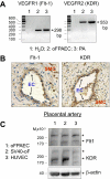
Figure 1
Figure 1
Expression of VEGFR1 and VEGFR2 in oFPAE cells ex vivo and in vitro. A, RT-PCR analysis of mRNA expression of VEGFRs in oFPAE cells and ovine fetoplacental arteries (PA). Total RNA (2 μg) sample isolated from oFPAE cells or homogenates of fetoplacental arteries of late pregnant (D120–130) ewes were reversed transcribed (RT), and PCR was carried out as described previously (28). The same PCR using total RNA from oFPAE cells as the template was run in parallel to exclude genomic DNA contamination (No-RT). B, Localization of VEGFR proteins in ovine fetoplacental arteries. Ovine fetoplacental artery segments were collected from late pregnant (D120–130) ewes immediately after death. The tissue segments were fixed with 3.7% paraformadehyde, paraffin embedded, and 6-μm sections were cut for immunohistochemical analysis of both VEGFR1 and VEGFR2 proteins with specific anti-Flt-1 pAb (5 μg/ml) (Santa Cruz) and anti-KDR pAb (5 μg/ml) (Upstate) by using the picture Plus Kit (Zymed Laboratories, Inc; South San Francisco, CA). Negative controls run in parallel with antirabbit IgG (5 μg/ml) displayed very weak or no brown color staining (data not shown). SMC, Smooth muscle cells. C, Western blot analysis of both VEGFR1/Flt1 and VEGFR2/KDR cellular protein extracts of oFPAE, SV40-oF, and HUVEC cells. Total cell extracts (20 μg/lane) were separated on 8% SDS-PAGE and transferred onto polyvinylidine difluoride membrane. Respective antibodies detect distinct bands of expected sizes of both VEGFR1 (Santa Cruz) and VEGFR2 (Upstate). Bands of expected sizes were indicated (arrowed). Asterisks indicate bands of 230–250 kDa that could be the glycosylated receptors. β-Actin serves as loading control.
Figure 2
Figure 2
Tyrosine phosphorylation of VEGFRs and their role in ERK2/1 activation by VEGF. A and B, VEGF activates both Flt-1/KDR in oFPAE cells. Serum-starved subconfluent oFPAE cells were treated with or without VEGF (10 ng/ml, 5 min). Protein extracts (1 mg/sample) were subjected to immunoprecipitation (IP) by pY99 antibody. The IP samples were subjected to Western blot analysis using anti-Flt-1 (Santa Cruz) or anti-KDR (Upstate) antibodies. C, Inhibition of VEGFR tyrosine kinase activity with SU5416 blocked VEGF-induced ERK2/1 activation. Cells were pretreated with various concentrations of SU5416 for 1 h, followed by treatment with VEGF (10 ng/ml) for 5 min. Cell extracts were prepared and subjected to Western blot analysis using specific antibodies against total ERK2/1 or phosphorylated ERK2/1. IB, Immunoblotting; IgG-H, IgG heavy chain.
Figure 3
Figure 3
VEGFR1 is not responsible for ERK2/1 activation by VEGF in oFPAE cells. A, VEGF, but not PlGF, activates ERK2/1 in oFPAE cells. oFPAE cells were treated with VEGF (10 ng/ml) or PlGF (10 ng/ml) for 5 min. B, Flt1 neutralizing antibody (R&D Systems) failed to suppress VEGF-stimulated ERK2/1 activation. oFPAE cells were treated with VEGFR1 neutralizing antibody at indicated concentrations for 1 h before VEGF (10 ng/ml) treatment for 5 min. Cell extracts were prepared and subjected to Western blot analysis using specific antibodies against phosphorylated or total ERK2/1. Antibodies against phospho-AKT1 (Ser473), AKT1, and phospho-p38MAPK (Thr180/Tyr182) were from Cell Signaling (Beverly, MA). Antibody against p38MAPK was from Santa Cruz. Bars represent mean ± sd of three separate experiments. Different letters indicate that the difference is significant (P < 0.05). Ab, Antibody.
Figure 4
Figure 4
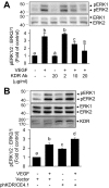
VEGFR2 is responsible for ERK2/1 activation by VEGF in oFPAE cells. A, KDR neutralizing antibody inhibited VEGF-stimulated ERK2/1 activation. oFPAE cells were treated with VEGFR2 neutralizing antibody (R&D Systems) at indicated concentrations for 1 h before VEGF (10 ng/ml) treatment for 5 min. B, KDR participates in ERK2/1 activation. oFPAE cells were transfected with phKDR/CE4.1 or vector plasmid 48 h before serum starvation and VEGF (10 ng/ml) treatment for 5 min. Cell extracts were prepared and subjected to Western blot analysis using antibodies against total or phosphorylated ERK2/1 or KDR (Upstate). Bars represent mean ± sd of three (A) or four (B) separate experiments. Different letters indicate that the difference is significant (P < 0.05). Ab, Antibody.
Figure 5
Figure 5
Role of ERK2/1 in the angiogenic responses to VEGF in oFPAE cells. A, U0126 suppresses VEGF-induced ERK2/1 activation. Serum-starved oFPAE cells were treated with U0126 at 5 or 10 μm for 1 h before VEGF (10 ng/ml) treatment for 5 min. Cell extracts were prepared and subjected to Western blot analysis using specific antibodies. B, Cell proliferation assay. Serum-starvated oFPAE cells were treated with or without VEGF (10 ng/ml) and/or U0126 (10 μm) as described in Materials and Methods. Bars represent mean ± sd of four separate determinations. C, Transwell migration assay. Migration was measured using 24-well Multiwell BD Falcon FluoroBlok Insert System (8.0-um pores, BD Biosciences) as described in Materials and Methods. The number of migrating cells was normalized to a 0.1% BSA control (Ctl). Bars represent mean ± sd of four separate determinations. D, Tube formation assays. Cultured oFPAE cells were seeded on growth factor-reduced Matrigel matrix in MCDB131 serum-free medium in the presence or absence of VEGF (10 ng/ml) and/or U0126 (10 μm) and incubated as described in Materials and Methods. Experiments were repeated four times independently, and statistical analyses were performed using Student’s t test. Bars with different letters differ significantly (P < 0.05).
Figure 6
Figure 6
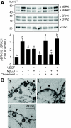
Integral caveolae are important for VEGF-stimulated ERK2/1 activation. A, Integrity of caveolae structure is important for VEGF-stimulated ERK2/1 activation in oFPAE cells. oFPAE cells were treated with or without a cholesterol depletant MβCD (10 mm) for 1 h, followed by with or without 0.2 mm of cholesterol repletion for 1 h, and then challenged with VEGF (10 ng/ml) for 5 min. Cell extracts were prepared and subjected to Western blot analysis. Bars (mean ± sd, n =4) with different letters differ significantly (P < 0.05). B, Electron microscopic analysis of the representative membrane structures of oFPAE cells under indicated treatments. a, oFPAE cells under resting condition; b, oFPAE cells treated with MβCD (10 mm, 1 h); c, oFPAE cells treated with MβCD (10 mm, 1 h) followed by cholesterol repletion (0.2 mm, 1 h). Arrows indicate the flask-shaped membranous invaginations (caveolae) on the membrane in a and c but absent in b. Cav1, Caveolin-1.
Figure 7
Figure 7
Down-regulation of caveolin-1 suppresses VEGF-induced angiogenic responses via ERK2/1 pathway. A, Establishment of stable oFPAE cells with down-regulated caveolin-1 expression. oFPAE cells transfected with caveolin-1 shRNAs (see Materials and Methods), selected with 600 μg/ml G418 in complete MCDB 131 medium. G418-resistant colonies were picked up and passaged. Down-regulation of caveolin-1 protein was verified by Western blot analysis. Note the clone from no. 1 shRNA transfection has greatest reduction of caveolin-1 protein (∼90%). Electron microscopic analysis showed that caveolin-1 down-regulation resulted in the loss of caveolae (arrows). B–D, Down-regulation of caveolin-1 inhibits VEGF-stimulated ERK2/1 activation (B), proliferation (C), and tube formation (D). Stable oFPAE cell lines harboring control and caveolin-1 shRNA were treated with VEGF for immunoblotting of total and phosphorylated ERK/2/1 (B), for proliferation (C), and for tube formation (D) assays, as described in Fig. 5. Bars (mean ± sd, n = 4) with different letters differ significantly (P < 0.05). Cav1, Caveolin-1; Ctl, control.
Figure 8
Figure 8
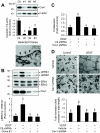
Overexpression of caveolin-1 suppresses VEGF-induced angiogenesis responses via ERK2/1 pathway. A and B, Overexpression of caveolin-1 with caveolin scaffolding domain (A) or caveolin-1 adenovirus (B) suppresses VEGF-induced ERK2/1 activation. oFPAE cells were treated with caveolin-scaffolding domain (Cav-SD) or its negative control (Cav-SD-X) at 5 μm for 2 h (A) or infected with caveolin-1 or GFP adenoviruses (5–10 MOI) for 24 h (B) before VEGF treatment (10 ng/ml, 5 min). Cell extracts were prepared and subjected to Western blot analysis using specific antibodies. C and D, Overexpression of caveolin-1 suppresses VEGF-stimulated oFPAE cell proliferation (C) and tube formation (D). oFPAE cells subjected to the proliferation (C) and tube formation (D) assays in the presence of Cav-SD or Cav-SD-X (5 μm) with or without VEGF (10 ng/ml). Bars (mean ± sd, n =4) with different letters differ significantly (P < 0.05). Cav1, Caveolin- 1; Ad-, adenovirus.
Figure 9
Figure 9
Subcellular fractionation of ERK2/1 signaling molecules in caveolae. A, Western blot analysis of fractions from sucrose-gradient centrifugation. SV40-oF cells were prepared as described in Materials and Methods and subjected to sucrose gradient centrifugation. Fractions (1 ml each) were collected from the top and analyzed by Western blotting with specific antibodies. B, Electron microscopic analysis of fraction 5. Note the isolated individual caveolae (arrows). C, Western blot analyses the ERK2/1 signaling components in fractions 5 and 9 with specific antibodies. Conc., Concentration; Fr#, fraction number; PLC, phospholipase C; S, sucrose; IgG-L, IgG light chain.
Figure 10
Figure 10
VEGF-stimulated ERK2/1 phosphorylation is regulated in caveolae. A, Time course of VEGF-induced ERK2/1 activation in caveolae. Serum-starved SV40-oF cells were treated with VEGF (10 ng/ml) for indicated times, prepared as described in Materials and Methods and subjected to sucrose gradient centrifugation. Fractions (1 ml each) were analyzed by Western blotting with specific antibodies. Note VEGF-induced ERK2/1 phosphorylation peaks in the caveolae around 5–15 min. Results are present as mean ± sd from four separate determinations. Asterisk indicates the difference is significant from control (P < 0.05). B, Integrity of caveolae structure is important for VEGF-stimulated ERK2/1 activation in caveolae. Serum-starved SV40-oF cells were treated with MβCD (10 mm, 1 h) and/or cholesterol repletion (0.2 mm, 1 h) as described in Fig. 6, prepared as described in Materials and Methods, and subjected to sucrose gradient centrifugation. Fractions (1 ml each) were analyzed by Western blotting with specific antibodies. Cav1, Caveolin-1.
Figure 11
Figure 11
VEGFR2 interacts with caveolin-1 in caveolae. A, KDR is localized in the caveolae. oFPAE cells were transfected with phKDR/CE4.1 or vector plasmid for 48 h before sucrose gradient centrifugation. Fractions (1 ml each) were analyzed by Western blotting with specific antibodies. B, Coimmunoprecipitation of KDR and caveolin-1. Protein extracts (1 mg/sample) of oFPAE cells transfected with phKDR/CE4.1 were immunoprecipitated (IP) with caveolin-1 or V5 antibody. The IP samples were subjected to Western blot analysis using anti-KDR (Upstate) or caveolin-1 antibody, respectively. C, Double immunofluorescence microscopy for subcellular localization of KDR and caveolin-1 in oFPAE cells. Cells were transfected with phKDR/CE4.1 for 48 h before labeling with both anti-V5 (green) and caveolin-1 (red) antibodies. Note colocalization of V5-taged KDR and caveolin-1 at the plasma membrane (arrows) of oFPAE cells. Abundant level of KDR is also detected in the cytoplasm of the cells, which could be due to the high level of expression of recombinant KDR. Conc., Concentration; IB, immunoblotting.

Similar articles

Cited by

References

    1. Reynolds LP, Redmer DA 2001 Angiogenesis in the placenta. Biol Reprod 64:1033–1040 - PubMed
    1. Magness RR, Rosenfeld CR 1989 The role of steroid hormones in the control of uterine blood flow. In: Rosenfeld CR, ed. Uterine circulation. Chap 10. Ithaca, NY: Perinatology Press; 239–271
    1. Cross JC, Werb Z, Fisher SJ 1994 Implantation and the placenta: key pieces of the development puzzle. Science 266:1508–1518 - PubMed
    1. Reynolds LP, Killilea SD, Redmer DA 1992 Angiogenesis in the female reproductive system. FASEB J 6:886–892 - PubMed
    1. Leach L, Babawale MO, Anderson M, Lammiman M 2002 Vasculogenesis, angiogenesis and the molecular organisation of endothelial junctions in the early human placenta. J Vasc Res 39:246–259 - PubMed

Publication types

Substances