

seeMotif: exploring and visualizing sequence motifs in 3D structures
- PMID: 19477961
- PMCID: PMC2703912
- DOI: 10.1093/nar/gkp439
seeMotif: exploring and visualizing sequence motifs in 3D structures
Abstract
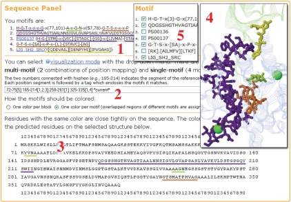
Sequence motifs are important in the study of molecular biology. Motif discovery tools efficiently deliver many function related signatures of proteins and largely facilitate sequence annotation. As increasing numbers of motifs are detected experimentally or predicted computationally, characterizing the functional roles of motifs and identifying the potential synergetic relationships between them are important next steps. A good way to investigate novel motifs is to utilize the abundant 3D structures that have also been accumulated at an astounding rate in recent years. This article reports the development of the web service seeMotif, which provides users with an interactive interface for visualizing sequence motifs on protein structures from the Protein Data Bank (PDB). Researchers can quickly see the locations and conformation of multiple motifs among a number of related structures simultaneously. Considering the fact that PDB sequences are usually shorter than those in sequence databases and/or may have missing residues, seeMotif has two complementary approaches for selecting structures and mapping motifs to protein chains in structures. As more and more structures belonging to previously uncharacterized protein families become available, combining sequence and structure information gives good opportunities to facilitate understanding of protein functions in large-scale genome projects. Available at: http://seemotif.csie.ntu.edu.tw,http://seemotif.ee.ncku.edu.tw or http://seemotif.csbb.ntu.edu.tw.
Figures

Similar articles
-
DBD2BS: connecting a DNA-binding protein with its binding sites.Nucleic Acids Res. 2012 Jul;40(Web Server issue):W173-9. doi: 10.1093/nar/gks564. Epub 2012 Jun 11. Nucleic Acids Res. 2012. PMID: 22693214 Free PMC article.
-
3MOTIF: visualizing conserved protein sequence motifs in the protein structure database.Bioinformatics. 2003 Mar 1;19(4):541-2. doi: 10.1093/bioinformatics/btf862. Bioinformatics. 2003. PMID: 12611811
-
MSDmotif: exploring protein sites and motifs.BMC Bioinformatics. 2008 Jul 17;9:312. doi: 10.1186/1471-2105-9-312. BMC Bioinformatics. 2008. PMID: 18637174 Free PMC article.
-
E1DS: catalytic site prediction based on 1D signatures of concurrent conservation.Nucleic Acids Res. 2008 Jul 1;36(Web Server issue):W291-6. doi: 10.1093/nar/gkn324. Epub 2008 Jun 4. Nucleic Acids Res. 2008. PMID: 18524800 Free PMC article.
-
Tools and resources for identifying protein families, domains and motifs.Genome Biol. 2002;3(1):REVIEWS2001. doi: 10.1186/gb-2001-3-1-reviews2001. Epub 2001 Dec 19. Genome Biol. 2002. PMID: 11806833 Free PMC article. Review.
References
Publication types
MeSH terms
LinkOut - more resources
Full Text Sources
Research Materials
