. 2009 Sep 1;83(4):688-97.
doi: 10.1093/cvr/cvp164. Epub 2009 May 28.

Cardiac fibroblast paracrine factors alter impulse conduction and ion channel expression of neonatal rat cardiomyocytes

Affiliations

Cardiac fibroblast paracrine factors alter impulse conduction and ion channel expression of neonatal rat cardiomyocytes

Dawn M Pedrotty et al. Cardiovasc Res. .

Abstract

Aims: The pathological proliferation of cardiac fibroblasts (CFs) in response to heart injury results in fibrosis, which correlates with arrhythmia generation and heart failure. Here we systematically examined the effect of fibroblast-derived paracrine factors on electrical propagation in cardiomyocytes.

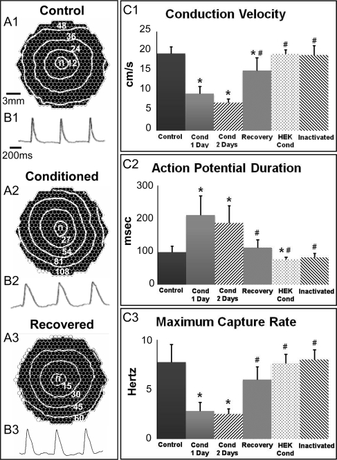
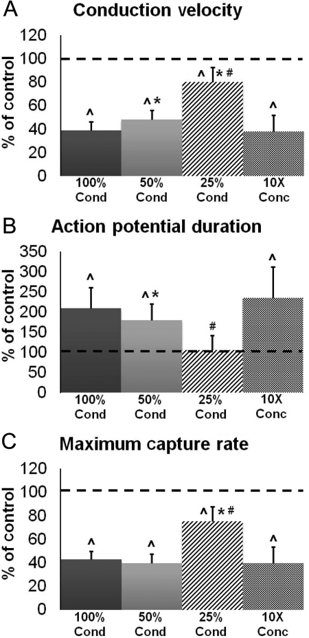
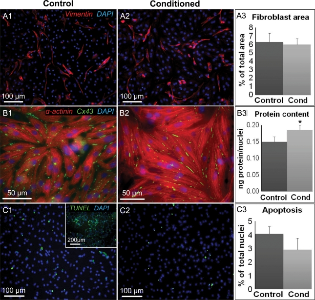
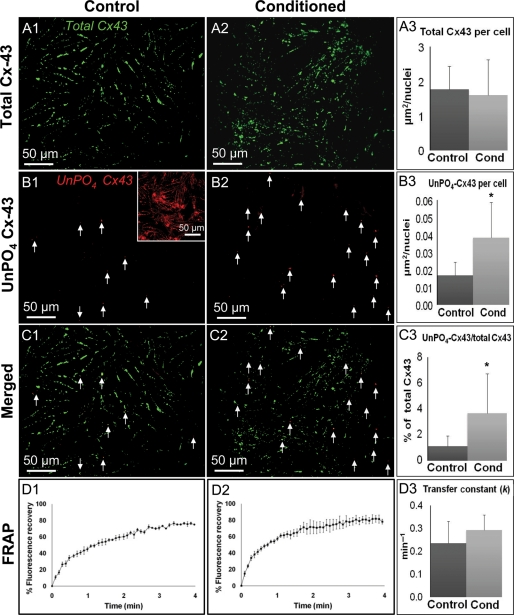
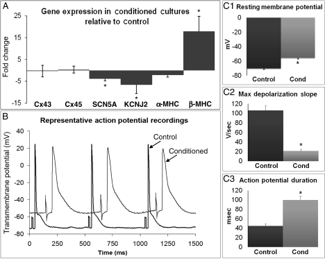
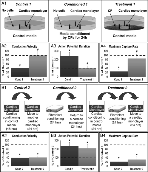
Methods and results: Neonatal rat cardiac monolayers were exposed for 24 h to media conditioned by CFs. Optical mapping, sharp microelectrode recordings, quantitative RT-PCR, and immunostaining were used to assess the changes in the propagation and shape of the action potential and underlying changes in gene and protein expression. The fibroblast paracrine factors produced a 52% reduction in cardiac conduction velocity, a 217% prolongation of action potential duration, a 64% decrease of maximum capture rate, a 21% increase in membrane resting potential, and an 80% decrease of action potential upstroke velocity. These effects were dose dependent and partially reversible with removal of the conditioned media. No fibroblast proliferation, cardiomyocyte apoptosis, or decreased connexin-43 expression, phosphorylation, and function were found in conditioned cardiac cultures. In contrast, the expression of the fast sodium, inward rectifying potassium, and transient outward potassium channels were, respectively, reduced 3.8-, 6.6-fold, and to undetectable levels. The expression of beta-myosin heavy chain increased 17.4-fold. No electrophysiological changes were observed from media conditioned by CFs in the presence of cardiomyocytes.

Conclusion: Paracrine factors from neonatal CFs alone produced significant electrophysiological changes in neonatal rat cardiomyocytes resembling those found in several cardiac pathologies.


Figures

Figure 1
Figure 1
Effect of CF-conditioned media on electrical propagation in cardiac monolayers. (A) Representative isochrone maps of control, CF-conditioned, and recovered cardiac monolayers stimulated by a point electrode (Π) at 2 Hz pacing rate. Recovered monolayers were exposed to CF-conditioned media for 24 h and then switched to control media for 24 h to recover. The isochrone lines are labelled in milliseconds. CVs were 19 cm/s (control), 8 cm/s (conditioned), and 15 cm/s (recovered). Circles represent 504 optical recording sites. (B) Optical action potential traces from respective monolayers in (A). (C) Parameters of electrical propagation. Cond, conditioned. Bars from left to right, respectively, correspond to n = 53, 53, 10, 10, 8, and 3 monolayers. Asterisks denote significantly different results from control; hash symbols indicate significantly different results from 1 and 2 day conditioned cultures.
Figure 2
Figure 2
Effect of CF-conditioned media dilution on electrical propagation in cardiac monolayers. Conditioned media (100% Cond) were diluted twice (50% Cond) and four times (25% Cond) by mixing with control media. CF paracrine factors in conditioned media were also concentrated 10× and diluted back to 1× using control media (10× Conc group). Electrophysiological parameters in (AC) are reported as per cent relative to unconditioned control (dashed line). Bars from left to right, respectively, correspond to n = 10, 10, 10, and 7 monolayers. Hat symbols denote significantly different results from control; asterisks denote significantly different results from 100% Cond; hash symbols indicate significantly different results from 50% Cond.
Figure 3
Figure 3
Effect of CF-conditioned media on cellular content, size, and apoptosis in cardiac monolayers. (A) Representative stainings for vimentin-positive fibroblasts in control (A1) and conditioned (A2) monolayers. (A3) Fibroblast percent area derived from the total of n = 4 monolayers per group. (B) Representative stainings showing cell size in control (B1) and conditioned (B2) monolayers. (B3) Total protein divided by number of nuclei was used as an index of cell size; n = 4 monolayers per group. (C) Representative stainings of apoptotic cells in control (C1) and conditioned (C2) monolayers. The inset in C1 is a positive control supplied by the manufacturer. (C3) Per cent of apoptotic nuclei derived from the total of n = 4 monolayers per group. Asterisks indicate significantly different results from control.
Figure 4
Figure 4
Effects of CF-conditioned media on the presence and phosphorylation of Cx43 in cardiac monolayers. (A) Representative stainings of total Cx43 in control (A1) and conditioned (A2) monolayers. (A3) Total Cx43 area per nucleus quantified from n = 5 monolayers per group. (B) Representative stainings of UnPO4 Cx43 in control (B1) and conditioned (B2) monolayers. Inset, positive control in hypoxic cardiomyocytes. Arrows point to positive staining (red). (B3) UnPO4 Cx-43 area per nucleus quantified from n = 5 monolayers per group. (C) Merged images of control (C1) and conditioned (C2) monolayers. (C3) The ratio of UnPO4-Cx43/total Cx43. (D) Time course of FRAP in control (D1) and conditioned (D2) cells averaged from n = 4 cells per group. (D3) The resulting transfer constants (k). Asterisks denote significantly different results from control.
Figure 5
Figure 5
Effects of CF-conditioned media on cardiac gene expression and single cell electrophysiology. (A) Gene expression in conditioned relative to control cultures. Cx, connexin; SCN5A, cardiac sodium channel NaV1.5 α-subunit; KCNJ2, inwardly rectifying potassium channel (Kir2.1); MHC, myosin heavy chain; KCND3, transient outward potassium channel (Kv4.3) was downregulated to an undetectable level by conditioning. (B) Representative cardiac action potentials traces from 2 Hz paced conditioned and control monolayers. (C13) Action potential parameters obtained from n = 18 (control) and 39 (conditioned) cells. Asterisks denote significantly different results from control.
Figure 6
Figure 6
Effects of CF-conditioning in the presence of cardiomyocytes or cardiac conditioned media. (A1) Experimental setup. Control 1, a cardiac monolayer exposed to control medium for 24 h. Conditioned 1 (Cond 1), a cardiac monolayer exposed to CF-conditioned media for 24 h. Treatment 1, a cardiac monolayer surrounded by cardiac fibroblasts (without direct contact) and exposed to control media for 24 h. (A2A4) Electrophysiological parameters in Cond 1 and Treatment 1, shown relative to Control 1 (dashed line) group. n = 12 monolayers per group. (B1) Experimental setup. Control 2, a cardiac monolayer exposed for 24 h to media conditioned by another cardiac monolayer for 48 h. Conditioned 2 (Cond 2), the same as Condition 1 in A1. Treatment 2, a cardiac monolayer exposed for 24 h to media conditioned for 24 h by CFs after being conditioned for 24 h by a cardiac monolayer. (B2–B4) Electrophysiological parameters in Cond 2 and Treatment 2, shown relative to Control 2 (dashed line) group. n = 12 monolayers per group. Asterisks denote significantly different results from control, while hash symbols denote significantly different from conditioned.

Similar articles

Cited by

References

    1. Camelliti P, Borg TK, Kohl P. Structural and functional characterisation of cardiac fibroblasts. Cardiovasc Res. 2005;65:40–51. - PubMed
    1. Nag A. Study of non-muscle cells of the adult mammalian heart: a fine structural analysis and distribution. Cytobios. 1980;28:41–61. - PubMed
    1. Banerjee I, Yekkala K, Borg TK, Baudino TA. Dynamic interactions between myocytes, fibroblasts, and extracellular matrix. Ann N Y Acad Sci. 2006;1080:76–84. - PubMed
    1. Brown R, Ambler S, Mitchell M, Long C. The cardiac fibroblast: therapeutic target in myocardial remodeling and failure. Annu Rev Pharmacol Toxicol. 2005;45:657–687. - PubMed
    1. de Bakker J, van Capelle F, Janse M, Tasseron S, Vermeulen J, de Jonge N, et al. Slow conduction in the infarcted human heart. ‘Zigzag’ course of activation. Circulation. 1993;88:915–926. - PubMed

Publication types

MeSH terms