Repeated maximal volitional effort contractions in human spinal cord injury: initial torque increases and reduced fatigue
- PMID: 19478056
- PMCID: PMC5603074
- DOI: 10.1177/1545968309336147
Repeated maximal volitional effort contractions in human spinal cord injury: initial torque increases and reduced fatigue
Abstract
Background: Substantial data indicate greater muscle fatigue in individuals with spinal cord injury (SCI) compared with healthy control subjects when tested by using electrical stimulation protocols. Few studies have investigated the extent of volitional fatigue in motor incomplete SCI.
Methods: Repeated, maximal volitional effort (MVE) isometric contractions of the knee extensors (KE) were performed in 14 subjects with a motor incomplete SCI and in 10 intact subjects. Subjects performed 20 repeated, intermittent MVEs (5 seconds contraction/5 seconds rest) with KE torques and thigh electromyographic (EMG) activity recorded.
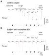
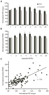
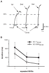
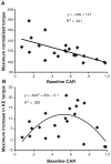
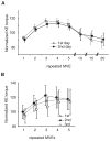
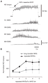
Results: Peak KE torques declined to 64% of baseline MVEs with repeated efforts in control subjects. Conversely, subjects with SCI increased peak torques during the first 5 contractions by 15%, with little evidence of fatigue after 20 repeated efforts. Increases in peak KE torques and the rate of torque increase during the first 5 contractions were attributed primarily to increases in quadriceps EMG activity, but not to decreased knee flexor co-activation. The observed initial increases in peak torque were dependent on the subject's volitional activation and were consistent on the same or different days, indicating little contribution of learning or accommodation to the testing conditions. Sustained MVEs did not elicit substantial increases in peak KE torques as compared to repeated intermittent efforts.
Conclusions: These data revealed a marked divergence from expected results of increased fatigability in subjects with SCI, and may be a result of complex interactions between mechanisms underlying spastic motor activity and changes in intrinsic motoneuron properties.
Figures
Similar articles
-
Central excitability contributes to supramaximal volitional contractions in human incomplete spinal cord injury.J Physiol. 2011 Aug 1;589(Pt 15):3739-52. doi: 10.1113/jphysiol.2011.212233. Epub 2011 May 24. J Physiol. 2011. PMID: 21610138 Free PMC article.
-
Muscle activation varies with contraction mode in human spinal cord injury.Muscle Nerve. 2015 Feb;51(2):235-45. doi: 10.1002/mus.24285. Epub 2014 Nov 19. Muscle Nerve. 2015. PMID: 24825184
-
The role of motor unit rate modulation versus recruitment in repeated submaximal voluntary contractions performed by control and spinal cord injured subjects.J Electromyogr Kinesiol. 2001 Jun;11(3):217-29. doi: 10.1016/s1050-6411(00)00055-9. J Electromyogr Kinesiol. 2001. PMID: 11335152
-
H-reflex, muscle voluntary activation level, and fatigue index of flexor carpi radialis in individuals with incomplete cervical cord injury.Neurorehabil Neural Repair. 2012 Jan;26(1):68-75. doi: 10.1177/1545968311418785. Epub 2011 Sep 27. Neurorehabil Neural Repair. 2012. PMID: 21952197
-
Short-term maximal-intensity resistance training increases volitional function and strength in chronic incomplete spinal cord injury: a pilot study.J Neurol Phys Ther. 2013 Sep;37(3):112-7. doi: 10.1097/NPT.0b013e31828390a1. J Neurol Phys Ther. 2013. PMID: 23673372
Cited by
-
Metabolic and Fatigue Profiles Are Comparable Between Prepubertal Children and Well-Trained Adult Endurance Athletes.Front Physiol. 2018 Apr 24;9:387. doi: 10.3389/fphys.2018.00387. eCollection 2018. Front Physiol. 2018. PMID: 29740332 Free PMC article.
-
Divergent modulation of clinical measures of volitional and reflexive motor behaviors following serotonergic medications in human incomplete spinal cord injury.J Neurotrauma. 2013 Mar 15;30(6):498-502. doi: 10.1089/neu.2012.2515. Epub 2013 Apr 3. J Neurotrauma. 2013. PMID: 22994901 Free PMC article. Clinical Trial.
-
Task-Specific Versus Impairment-Based Training on Locomotor Performance in Individuals With Chronic Spinal Cord Injury: A Randomized Crossover Study.Neurorehabil Neural Repair. 2020 Jul;34(7):627-639. doi: 10.1177/1545968320927384. Epub 2020 Jun 1. Neurorehabil Neural Repair. 2020. PMID: 32476619 Free PMC article. Clinical Trial.
-
Increased spinal reflex excitability is associated with enhanced central activation during voluntary lengthening contractions in human spinal cord injury.J Neurophysiol. 2015 Jul;114(1):427-39. doi: 10.1152/jn.01074.2014. Epub 2015 May 13. J Neurophysiol. 2015. PMID: 25972590 Free PMC article.
-
Repeated and patterned stimulation of cutaneous reflex pathways amplifies spinal cord excitability.J Neurophysiol. 2020 Aug 1;124(2):342-351. doi: 10.1152/jn.00072.2020. Epub 2020 Jun 24. J Neurophysiol. 2020. PMID: 32579412 Free PMC article.
References
-
- Castro MJ, Apple DF, Jr, Staron RS, et al. Influence of complete spinal cord injury on skeletal muscle within 6 mo of injury. J Appl Physiol. 1999;86:350–358. - PubMed
-
- Shields RK. Fatigability, relaxation properties, and electromyographic responses of the human paralyzed soleus muscle. J Neurophysiol. 1995;73:2195–2206. - PubMed
-
- Crameri RM, Weston AR, Rutkowski S, et al. Effects of electrical stimulation leg training during the acute phase of spinal cord injury: a pilot study. Eur J Appl Physiol. 2000;83:409–415. - PubMed
-
- Talmadge RJ, Castro MJ, Apple DF, Jr, et al. Phenotypic adaptations in human muscle fibers 6 and 24 wk after spinal cord injury. J Appl Physiol. 2002;92:147–154. - PubMed
-
- Olive JL, Dudley GA, McCully KK. Vascular remodeling after spinal cord injury. Med Sci Sports Exerc. 2003;35:901–907. - PubMed
Publication types
MeSH terms
Grants and funding
LinkOut - more resources
Full Text Sources
Medical

