. 2009 Jun 9;106(23):9286-91.
doi: 10.1073/pnas.0900668106. Epub 2009 May 28.

Stem cells are dispensable for lung homeostasis but restore airways after injury

Affiliations

Stem cells are dispensable for lung homeostasis but restore airways after injury

Adam Giangreco et al. Proc Natl Acad Sci U S A. .

Abstract

Local tissue stem cells have been described in airways of the lung but their contribution to normal epithelial maintenance is currently unknown. We therefore developed aggregation chimera mice and a whole-lung imaging method to determine the relative contributions of progenitor (Clara) and bronchiolar stem cells to epithelial maintenance and repair. In normal and moderately injured airways chimeric patches were small in size and not associated with previously described stem cell niches. This finding suggested that single, randomly distributed progenitor cells maintain normal epithelial homeostasis. In contrast we found that repair following severe lung injury resulted in the generation of rare, large clonal cell patches that were associated with stem cell niches. This study provides evidence that epithelial stem cells are dispensable for normal airway homeostasis. We also demonstrate that stem cell activation and robust clonal cellular expansion occur only during repair from severe lung injury.


Conflict of interest statement

Conflict of interest: The authors declare no conflict of interest.

Figures

Fig. 1.
Fig. 1.
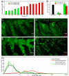
Chimeric epithelial patches do not associate with airway stem cell niches. (A and B) Representative low (A) and high magnification (B) lung whole mount immunostained for GFP (green) to reveal the distribution of chimeric epithelial patches. (C and D) High-magnification images reveal that CGRP-immunoreactive NEBs (red cell cluster, C) and BADJ-associated stem cell niches (arrow, D) do not correlate with GFP-expressing patches. (E) Quantification of chimeric patch correlation with defined airway stem cells. (F and G) Tissue sections stained for CCSP, CGRP, and GFP to visualize GFP chimeric patch distribution with respect to NEB (red, F) and BADJ stem cell niches (right side, G). (H and I) BADJ tissue staining for CCSP (blue), SPC (red), and GFP (green) to compare CCSP/SPC dual-positive cell chimerism (arrows) with adjacent alveolar septae. The proximal–distal axis (PD) and airway boundaries (white lines) are indicated in A and D. (Scale bars, 50 μm.)
Fig. 2.
Fig. 2.
Epithelial patch size suggests that airways are not maintained by stem cells. (A and B) Expected GFP patch size and abundance if stem (A) or single progenitor cells (B) maintain conducting airways. (C and D) Images of GFP-expressing cell patches within airways before (C) and after image pseudocoloring (D). (Scale bars: C and D, 50 μm.) (E) Overall GFP expression within airways of 10 individual mice. (F) Representative pseudocolored chimeric airway used for airway patch size quantification. (G) Analysis comparing GFP-positive and -negative patch size vs. frequency among the lowest (mouse A, black line), highest (mouse J, green line), and average of all chimeric airways (red line). Data represent mean ± SEM (G). PD indicates proximal–distal axis in F.
Fig. 3.
Fig. 3.
Chimeric patches are proliferation competent and multipotent. (A) Immunostaining for GFP (green), CCSP (blue), and Ki67 (red) to identify GFP(+) and -(−) mitotic cells (arrows). (B and C) Immunofluorescent detection of BrdU (red) plus CGRP (green, B) or CCSP (green, C). White arrows indicate mitotic cells; the yellow line (B) and arrow (C) denote the NEB and BADJ. (D) Whole-mount airway immunostaining for GFP (green) plus CCSP (red) to identify multipotent epithelial patches. (E) Tissue CCSP, ACT, and GFP immunostaining reveals GFP-expressing ciliated cells (arrows). (F) Tissue CCSP, CGRP, and GFP immunostaining identifies intrapulmonary neuroepithelial bodies containing GFP(+) and -(−) neuroendocrine cells (white and yellow arrows). The white line denotes epithelial basement membrane (A–C, E, and F). (Scale bars, 50 μm.)
Fig. 4.
Fig. 4.
Injury severity determines epithelial repair modality. (A) Percentage of body weight loss 48 h postnaphthalene administration. Individual chimeric mice were clustered on the basis of low (<10%, green) or high weight loss (>10%, red). (B) Lavageable CCSP protein abundance in control, 3-day recovered, or 30-day recovered animals. (C–F) Representative images of chimeric epithelial patches within 3-day (C and D) and 30-day (E and F) recovered airways grouped according to slight (C and E) or severe (D and F) injury/48-h weight loss. (Scale bars: C–F, 100 μm.) (G) Airway chimeric patch size-frequency plots comparing uninjured (black line), 3- or 30-day slight (dashed and solid green lines), and 30-day severe injury airways (red line). The proximal–distal axes (PD) and airway boundaries (white lines) are indicated in C–F. Data are represented as mean ± SEM in B and G. We used Volocity image acquisition software to normalize the brightness and contrast on regions acquired during an intermittent laser anomaly.
Fig. 5.
Fig. 5.
NEB- and BADJ-associated stem cells define regenerating patches within severe injury airways. (A–C) CGRP (red), GFP (green), and CCSP (blue) immunostaining identifies NEB (red cell clusters, A and B) and BADJ (arrows, C) -associated epithelial regeneration 30 days post injury. (D) Immunostaining also identified numerous non-branchpoint-associated NEBs that lacked associated regeneration. (E) Frequency of regenerative vs. nonregenerative NEBs. (F and G) Tissue section staining confirms epithelial regeneration adjacent to branchpoint-associated NEBs (red cell cluster, F) and terminal bronchioles (arrow, G). (H) Model in which epithelial injury severity is the principal determinant of progenitor cell-dependent homeostasis or stem cell-mediated epithelial repair. Data are represented as mean ± SEM. (Scale bars, 50 μm.)

References

    1. Rawlins EL, Hogan BL. Ciliated epithelial cell lifespan in the mouse trachea and lung. Am J Physiol. 2008;295:L231–L234. - PMC - PubMed
    1. Rawlins EL, Ostrowski LE, Randell SH, Hogan BL. Lung development and repair: Contribution of the ciliated lineage. Proc Natl Acad Sci USA. 2007;104:410–417. - PMC - PubMed
    1. Stripp BR, Reynolds SD. Maintenance and repair of the bronchiolar epithelium. Proc Am Thorac Soc. 2008;5:328–333. - PMC - PubMed
    1. Evans MJ, Cabral-Anderson LJ, Freeman G. Role of the Clara cell in renewal of the bronchiolar epithelium. Lab Invest. 1978;38:648–653. - PubMed
    1. Dor Y, Brown J, Martinez OI, Melton DA. Adult pancreatic beta-cells are formed by self-duplication rather than stem-cell differentiation. Nature. 2004;429:41–46. - PubMed

Publication types