. 2009 Jun 9;106(23):9373-8.
doi: 10.1073/pnas.0903487106. Epub 2009 May 28.

Unphosphorylated STAT1 prolongs the expression of interferon-induced immune regulatory genes

Affiliations

Unphosphorylated STAT1 prolongs the expression of interferon-induced immune regulatory genes

Hyeonjoo Cheon et al. Proc Natl Acad Sci U S A. .

Abstract

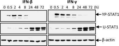
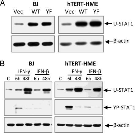
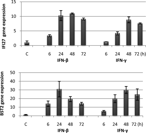
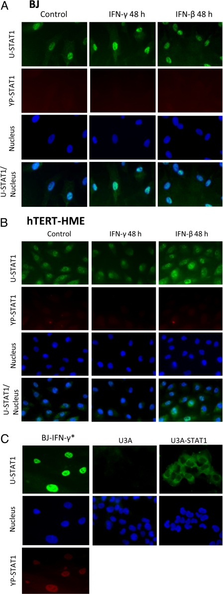
In normal human cells treated with interferons (IFNs), the concentration of tyrosine-phosphorylated STAT1 (YP-STAT1), which drives the expression of a large number of genes, increases quickly but then decreases over a period of several hours. Because the STAT1 gene is activated by YP-STAT1, IFNs stimulate a large increase in the concentration of unphosphorylated STAT1 (U-STAT1) that persists for several days. To test the significance of high U-STAT1 expression, we increased its concentration exogenously in the absence of IFN treatment. In response, the expression of many immune regulatory genes (e.g., IFI27, IFI44, OAS, and BST2) was increased. In human fibroblasts or mammary epithelial cells treated with low concentrations of IFN-beta or IFN-gamma, the expression of the same genes increased after 6 h and continued to increase after 48 or 72 h, long after the concentration of YP-STAT1 had returned to basal levels. Consistent with its activity as a transcription factor, most U-STAT1 was present in the nuclei of these cells before IFN treatment, and the fraction in nuclei increased 48 h after treatment with IFN. We conclude that the nuclear U-STAT1 that accumulates in response to IFNs maintains or increases the expression of a subset of IFN-induced genes independently of YP-STAT1, and that many of the induced proteins are involved in immune regulation.


Conflict of interest statement

The authors declare no conflict of interest.

Figures

Fig. 1.
Fig. 1.
IFN-β or IFN-γ increases the expression of U-STAT1. hTERT-HME1 cells were treated with IFN-β (50 units/mL) or IFN-γ (1 ng/mL). The amounts of YP-STAT1 and U-STAT1 were measured by the Western blotting method.
Fig. 2.
Fig. 2.
The expression of U-STAT1 and YP-STAT1 in cells used in microarray analyses. (A) hTERT-HME1 or BJ cells were infected with lentiviruses expressing wild-type (WT) or Y701F-STAT1 (YF), or with empty vector (Vec). The U-STAT1 protein expression levels were measured by the Western blotting method. (B) Cells were treated with IFN-β or IFN-γ for 6 or 48 h to establish a condition in which the maximum levels of U-STAT1 were induced at 48 h, with the minimum levels of YP-STAT1. The selected concentrations of IFNs (0.3 ng/mL IFN-γ or 3 units/mL IFN-β for BJ cells, and 0.1 ng/mL IFN-γ or 5 units/mL IFN-β for hTERT-HME1 cells) were applied, and the amounts of U-STAT1 and PY-STAT1 were analyzed by the Western blotting method.
Fig. 3.
Fig. 3.
U-STAT1 prolongs the expression of some IFN-induced immune regulatory genes. BJ cells were treated with IFN-β (3 units/mL) or IFN-γ (0.3 units/mL), and the expression of IFI27 and BST2 was measured by real-time PCR. The figure shows the mean values, with standard deviations, of triplicate experiments.
Fig. 4.
Fig. 4.
Most U-STAT1 is located in nuclei of untreated and IFN-treated BJ or hTERT-HME1 cells. (A) BJ cells were treated with 0.3 ng/mL IFN-γ or 3 units/mL IFN-β for 48 h. Immunocytochemistry was performed with antibodies against U-STAT1 (green) or YP-STAT1 (red). Nuclei were stained with DAPI. YP-STAT1 was not seen in untreated cells or in cells treated with IFNs for 48 h. (B) hTERT-HME1 cells were treated with 0.1 ng/mL IFN-γ or 5 units/mL IFN-β for 48 h. YP-STAT1 was not seen in untreated cells or in cells treated with IFNs for 48 h. (C) As a positive control for PY-STAT1 staining, cells were treated with 3 ng/mL IFN-γ (IFN-γ*) for 1 h, which shows PY-STAT1 staining inside nuclei. U3A cells (STAT1-null 2fTGH cells) were used as a negative control, with same antibodies used in A and B. Untreated U3A cells transfected with wild-type STAT1 show clear cytoplasmic localization of U-STAT. The magnification of the images is ×400.

Similar articles

Cited by

References

    1. Stark GR, Kerr IM, Williams BR, Silverman RH, Schreiber RD. How cells respond to interferons. Annu Rev Biochem. 1998;67:227–264. - PubMed
    1. Stark GR. How cells respond to interferons revisited: From early history to current complexity. Cytokine Growth Factor Rev. 2007;18:419–423. - PMC - PubMed
    1. Uddin S, et al. Activation of the p38 mitogen-activated protein kinase by type I interferons. J Biol Chem. 1999;274:30127–30131. - PubMed
    1. Sizemore N, et al. Inhibitor of κB kinase is required to activate a subset of interferon γ-stimulated genes. Proc Natl Acad Sci USA. 2004;101:7994–7998. - PMC - PubMed
    1. Qing Y, Stark GR. Alternative activation of STAT1 and STAT3 in response to interferon-γ. J Biol Chem. 2004;279:41679–41685. - PubMed