. 2009 Jul 24;284(30):20079-89.
doi: 10.1074/jbc.M109.018010. Epub 2009 May 28.

Multiple conformers in active site of human dihydrofolate reductase F31R/Q35E double mutant suggest structural basis for methotrexate resistance

Affiliations

Multiple conformers in active site of human dihydrofolate reductase F31R/Q35E double mutant suggest structural basis for methotrexate resistance

Jordan P Volpato et al. J Biol Chem. .

Abstract

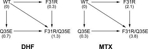
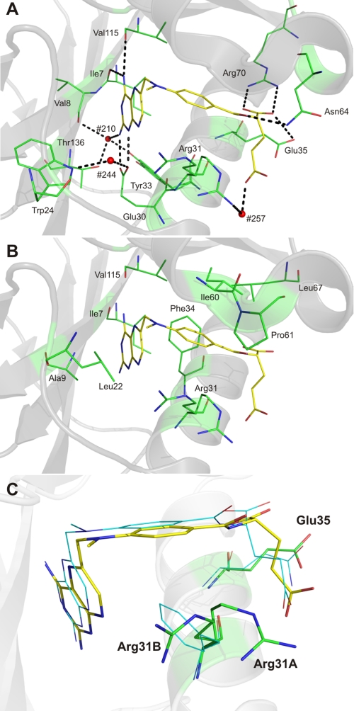
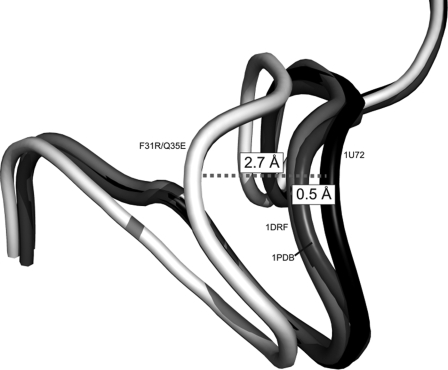
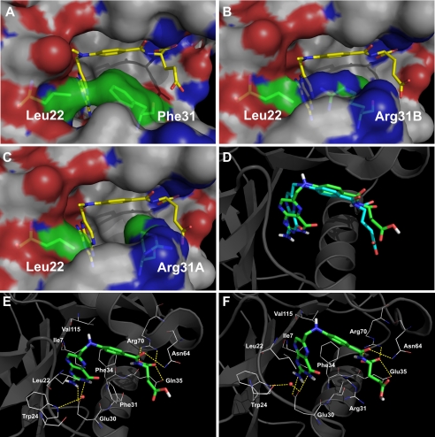
Methotrexate is a slow, tight-binding, competitive inhibitor of human dihydrofolate reductase (hDHFR), an enzyme that provides key metabolites for nucleotide biosynthesis. In an effort to better characterize ligand binding in drug resistance, we have previously engineered hDHFR variant F31R/Q35E. This variant displays a >650-fold decrease in methotrexate affinity, while maintaining catalytic activity comparable to the native enzyme. To elucidate the molecular basis of decreased methotrexate affinity in the doubly substituted variant, we determined kinetic and inhibitory parameters for the simple variants F31R and Q35E. This demonstrated that the important decrease of methotrexate affinity in variant F31R/Q35E is a result of synergistic effects of the combined substitutions. To better understand the structural cause of this synergy, we obtained the crystal structure of hDHFR variant F31R/Q35E complexed with methotrexate at 1.7-A resolution. The mutated residue Arg-31 was observed in multiple conformers. In addition, seven native active-site residues were observed in more than one conformation, which is not characteristic of the wild-type enzyme. This suggests that increased residue disorder underlies the observed methotrexate resistance. We observe a considerable loss of van der Waals and polar contacts with the p-aminobenzoic acid and glutamate moieties. The multiple conformers of Arg-31 further suggest that the amino acid substitutions may decrease the isomerization step required for tight binding of methotrexate. Molecular docking with folate corroborates this hypothesis.


Figures

FIGURE 1.
FIGURE 1.
Chemical structures of hDHFR ligands. Atom numbering is shown on DHF.
FIGURE 2.
FIGURE 2.
Double mutant cycle of F31R/Q35E for DHF and MTX affinity. Numbers in parentheses are ΔΔG values (in kilocalories/mol) for each variant relative to the WT, for KmDHF and for KiMTX, respectively.
FIGURE 3.
FIGURE 3.
Active site residues observed as two or more conformers in the crystal structure of the F31R/Q35E mutant. MTX is shown in line representation and relevant residues are shown as sticks, colored by atom (C: yellow (MTX), orange (conformers ‘a’), and cyan (conformers ‘b’); O: red; N: blue). Residues Ser-118, Thr-145, and Asp-146 belong to the NADPH-binding sub-site.
FIGURE 4.
FIGURE 4.
Bound MTX in hDHFR variant F31R/Q35E. Polar (A) and non-polar interactions (B). MTX is shown in stick representation, and relevant residues are shown as lines, colored by atom (C: yellow (MTX) and green (active-site residues); O: red; N: blue). In A, H-bonds and salt bridges are shown as dashed black lines, while active-site water molecules #210, #244, and #257 are shown as red spheres. The backbone carbonyls of Ile-7 and Val-115 are within H-bonding distance of the pterin 4-amino group, as is the hydroxyl group of Tyr-121. The carboxylate of the catalytic Glu-30 residue forms a salt bridge with the pterin N1 and 2-amino group. A conserved active-site water molecule (H2O #257), coordinated via H-bonding interactions with the indole ring of Trp-24 and the Glu-30 carboxylate group, is within H-bonding distance of the pterin N8. Another highly conserved water molecule (H2O #210) present in the active site can H-bond with the pterin 2-amino group, the backbone carbonyl of Val-8, and the hydroxyl group of Thr-136. The p-ABA moiety of MTX interacts mainly with residues Phe-34, Ile-60, Pro-61, and Leu-67 through hydrophobic contacts. A sole H-bond is formed between the carbonyl group of the p-ABA moiety and the γ-amino group of residue Asn-64. C, position of MTX, Arg-31, and Glu-35 in F31R/Q35E relative to the position observed in WT hDHFR (1U72). Residues and MTX from the F31R/Q35E structure are shown in stick representation, whereas residues and MTX from superposed WT hDHFR (1U72) are shown as lines, colored by atom (C: green (F31R/Q35E), cyan (WT hDHFR), and yellow (MTX from 3EIG); O: red; N: blue). Superposition was performed by Cα alignment of the crystal structures.
FIGURE 5.
FIGURE 5.
Shift of loop 17–27 in hDHFR variant F31R/Q35E. Loops are shown in schematic representation. Residues 17–27 are shown for variant F31R/Q35E (white), and WT hDHFR from three structures: 1U72 (complexed with NADPH and MTX; black), 1DRF (complexed with folate; light gray), and 1PDB (apoenzyme; dark gray). Distances were calculated between the 1U72 Cα of Gly-20 of 1U72 and the respective Cα of Gly-20 for the three structures. Superposition was performed by Cα alignment of the crystal structures.
FIGURE 6.
FIGURE 6.
Comparison of WT hDHFR and variant F31R/Q35E by modeling. A–C, surface representation of the contacts established between residues 22 and 31 in WT hDHFR (A, 1U72), variant F31R/Q35E with Arg-31B (B) or with Arg-31A (C) bound to MTX. MTX and residues 22 and 31 are in stick representation, colored by atom (C: yellow (MTX) and green (residues 22 and 31), O: red, N: blue). Surface is colored by atoms (C: white, O: red, N: blue). D–F, docking of folate onto WT hDHFR and variant F31R/Q35E. D, superposition of the original crystal structure 1DRF (WT DHFR bound to folate in blue) and the docking model of 1U72 (WT DHFR) docked with folate (in green). Results for the minimum energy binding conformers are shown for folate (E) docked onto WT hDHFR (1U72), as well as for folate (F) docked onto F31R/Q35E with Arg-31A conformer. The ligands are shown in stick representation while the residues are shown as lines, colored by atom (C: green (ligands) and white (residues), O: red, N: blue). Superposition was performed by Cα alignment of the crystal structures.

Similar articles

Cited by

References

    1. Slamon D. J., Romond E. H., Perez E. A. (2006) Clin. Adv. Hematol. Oncol. 4, Suppl. 1, 4–9 - PubMed
    1. Daw N. C., Billups C. A., Rodriguez-Galindo C., McCarville M. B., Rao B. N., Cain A. M., Jenkins J. J., Neel M. D., Meyer W. H. (2006) Cancer 106, 403–412 - PubMed
    1. Strojan P., Soba E., Budihna M., Auersperg M. (2005) J. Surg. Oncol. 92, 278–283 - PubMed
    1. Ramanan A. V., Whitworth P., Baildam E. M. (2003) Arch. Dis. Child 88, 197–200 - PMC - PubMed
    1. Flintoff W. F., Sadlish H., Gorlick R., Yang R., Williams F. M. (2004) Biochim. Biophys. Acta 1690, 110–117 - PubMed

Publication types