. 2009 Jul 31;284(31):21036-46.
doi: 10.1074/jbc.M109.016766. Epub 2009 May 28.

Occludin phosphorylation and ubiquitination regulate tight junction trafficking and vascular endothelial growth factor-induced permeability

Affiliations

Occludin phosphorylation and ubiquitination regulate tight junction trafficking and vascular endothelial growth factor-induced permeability

Tomoaki Murakami et al. J Biol Chem. .

Abstract

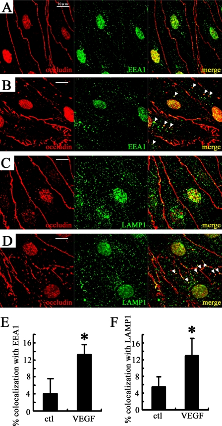
Vascular endothelial growth factor (VEGF) alters tight junctions (TJs) and promotes vascular permeability in many retinal and brain diseases. However, the molecular mechanisms of barrier regulation are poorly understood. Here we demonstrate that occludin phosphorylation and ubiquitination regulate VEGF-induced TJ protein trafficking and concomitant vascular permeability. VEGF treatment induced TJ fragmentation and occludin trafficking from the cell border to early and late endosomes, concomitant with increased occludin phosphorylation on Ser-490 and ubiquitination. Furthermore, both co-immunoprecipitation and immunocytochemistry demonstrated that VEGF treatment increased the interaction between occludin and modulators of intracellular trafficking that contain the ubiquitin interacting motif, including Epsin-1, epidermal growth factor receptor pathway substrate 15 (Eps15), and hepatocyte growth factor-regulated tyrosine kinase substrate (Hrs). Inhibiting occludin phosphorylation by mutating Ser-490 to Ala suppressed VEGF-induced ubiquitination, inhibited trafficking of TJ proteins, and prevented the increase in endothelial permeability. In addition, an occludin-ubiquitin chimera disrupted TJs and increased permeability without VEGF. These data demonstrate a novel mechanism of VEGF-induced occludin phosphorylation and ubiquitination that contributes to TJ trafficking and subsequent vascular permeability.


Figures

FIGURE 1.
FIGURE 1.
VEGF disrupts continuous cell border staining of occludin, and increases the co-localization between occludin and endosomal markers in BREC. A and B, double staining of occludin and early endosome antigen 1 (EEA1) in control (A) and after 15 min with 50 ng/ml VEGF treatment (B). C and D, immunolocalization of occludin and lysosome-associated membrane protein 1 (LAMP1) under control conditions (C) or after VEGF treatment (50 ng/ml, 15 min) (D). Arrowheads, puncta with co-localization. Background nuclear staining was also observed with secondary antibody alone (not shown). Scale bar = 10 μm. The co-localization was quantified (E and F). n = 5. Error bars represent the S.D. *, p < 0.01 versus control (ctl).
FIGURE 2.
FIGURE 2.
VEGF increases occludin ubiquitination, dependent on phosphorylation at Ser-490. A, BRECs treated with VEGF (50 ng/ml) over the time course indicated were used to detect the ubiquitination of occludin (A) and phospho-Ser-490 (B). Arrowheads, ubiquitinated occludin. C, occludin detected by rabbit occludin antibody (Zymed Laboratories Inc., 71–1500) and phospho-Ser-490 were also conjugated by exogenous ubiquitin after VEGF treatment (50 ng/ml for 15 min). D, VEGF treatment (50 ng/ml, 20 min) increased the ubiquitination of transfected WtOcc but not S490AOcc. HA-Ub, HA-tagged ubiquitin; WT, WtOcc; S490A, S490AOcc. IB, immunoblot; IP, immunoprecipitated.
FIGURE 3.
FIGURE 3.
VEGF induces the interaction between occludin and the E3 ligase, Itch, that requires occludin phosphorylation at Ser-490. A and B, BRECs were incubated with VEGF (50 ng/ml) for the indicated time points, and used for co-immunoprecipitation (IP) studies with anti-occludin (A) or anti-Itch (B) antibody. The interaction between occludin and Itch was increased in a time-dependent manner. C, after transfection, WtOcc or S490AOcc were co-immunoprecipitated. VEGF (50 ng/ml, 15 min) increased the interaction between WtOcc and Itch, which was inhibited by S490AOcc. WT, WtOcc; S490A, S490AOcc. IB, immunoblot.
FIGURE 4.
FIGURE 4.
VEGF promotes the interaction between occludin and Epsin-1 or Eps15. A, co-immunoprecipitation (IP) with antibodies against occludin, Eps15, or Epsin-1 was performed in the absence or presence of VEGF (50 ng/ml, 15 min). VEGF increased the protein-protein interaction between total occludin (or phospho-Ser-490) and Eps15 or Epsin-1. Occludin was detected by rabbit occludin antibody (Zymed Laboratories Inc., 71-1500). pS490, phospho-Ser-490. Immunocytochemistry in the absence (B and D) or presence (C and E) of VEGF (50 ng/ml, 15 min) was performed. Compared with baseline, both TJ disruption and co-localization with clathrin (arrowheads) were observed after VEGF treatment. Arrowheads, co-localization of cytoplasmic puncta. Scale bar = 10 μm. IB, immunoblot.
FIGURE 5.
FIGURE 5.
VEGF promotes the interaction between occludin and Hrs. Immunocytochemistry was performed on BREC without (A) or with (B) VEGF treatment (50 ng/ml, 15 min). At baseline, occludin did not colocalize with Hrs, whereas VEGF increased the intracellular punctate staining of occludin, which co-localized with Hrs (arrowheads). C, co-immunoprecipitation (IP) with anti-occludin or anti-Hrs antibody showed that the interaction was increased by VEGF. Occludin around 65 kDa was detected by rabbit occludin antibody (Zymed Laboratories Inc., 71-1500). pS490, phospho-Ser-490. Scale bar = 10 μm. IB, immunoblot.
FIGURE 6.
FIGURE 6.
S490AOcc mutant inhibits occludin trafficking. A, WtOcc or S490AOcc was expressed and co-immunoprecipitated (IP) with V5 antibody. VEGF (50 ng/ml, 15 min) induced the interaction between WtOcc and Epsin-1, Eps15, or Hrs, whereas S490AOcc completely inhibited these interactions. B, immunocytochemistry demonstrated that VEGF disrupted continuous occludin staining in WtOcc-transfected cells, whereas transfection of the S490AOcc mutant prevented the VEGF-induced TJ disruption. These changes were quantified and presented in supplemental Fig. S3. WT, WtOcc; S490A, S490AOcc. Scale bar = 10 μm.
FIGURE 7.
FIGURE 7.
Occludin-ubiquitin chimera induces occludin trafficking. A, co-immunoprecipitation (IP) demonstrated that both Occ-Ub and S490A-Ub interact with Epsin-1, Eps15, and Hrs in the absence of VEGF. B, BRECs were transfected with these chimeric mutants for immunocytochemistry. Both Occ-Ub and S490A-Ub presented intracellular puncta, and cell border staining of occludin was fragmented in Occ-Ub- or S490A-Ub-transfected cells. WT, WtOcc. Scale bar = 10 μm. IB, immunoblot.
FIGURE 8.
FIGURE 8.
VEGF-induced occludin degradation is mediated via the ubiquitin/proteasomal system. A, after protein synthesis was inhibited by cycloheximide (CHX) (10 μg/ml) occludin content was evaluated by Western blotting. VEGF (50 ng/ml) enhanced the rate of occludin degradation, which was blocked by MG132 (50 μm). The rate of claudin-5 degradation was not changed by either VEGF or MG132. n = 7–8. Error bars represent the S.D. B, after the pretreatment with MG132 (50 μm), BRECs were treated with VEGF (50 ng/ml, 15 min). MG132 increased ubiquitinated occludin with or without VEGF. IP, immunoprecipitated; IB, immunoblot.
FIGURE 9.
FIGURE 9.
Phosphorylation of occludin on Ser-490 and ubiquitination regulate VEGF-induced vascular endothelial permeability. A, diffusive flux (Po) of 70-kDa-dextran was evaluated in the absence or presence of VEGF (50 ng/ml), after the transfection of each occludin mutant. VEGF-induced hyperpermeability was inhibited by S490AOcc, whereas Occ-Ub and S490A-Ub increased the tracer flux without VEGF. B, transendothelial electrical resistance (TER) measurements. VEGF reduced the resistance, which was inhibited by S490AOcc. The chimera proteins decreased the resistance both with and without VEGF. n = 7–9. Error bars represent the S.D. * p < 0.01 versus EV-transfected cells without VEGF treatment. WT, WtOcc; S490A, S490AOcc. NS, not significant.

References

    1. Antonetti D. A., Barber A. J., Bronson S. K., Freeman W. M., Gardner T. W., Jefferson L. S., Kester M., Kimball S. R., Krady J. K., LaNoue K. F., Norbury C. C., Quinn P. G., Sandirasegarane L., Simpson I. A. (2006) Diabetes 55, 2401–2411 - PubMed
    1. Harhaj N. S., Antonetti D. A. (2004) Int. J. Biochem. Cell Biol. 36, 1206–1237 - PubMed
    1. Aiello L. P. (1997) Invest. Ophthalmol. Vis. Sci. 38, 1647–1652 - PubMed
    1. Machein M. R., Plate K. H. (2000) J. Neurooncol. 50, 109–120 - PubMed
    1. Gardner T. W., Antonetti D. A. (2008) Curr. Diab. Rep. 8, 263–269 - PubMed

Publication types

MeSH terms