. 2009 Jul;20(7):1491-503.
doi: 10.1681/ASN.2008101117. Epub 2009 May 28.

Slit diaphragms contain tight junction proteins

Affiliations

Slit diaphragms contain tight junction proteins

Hirotaka Fukasawa et al. J Am Soc Nephrol. 2009 Jul.

Abstract

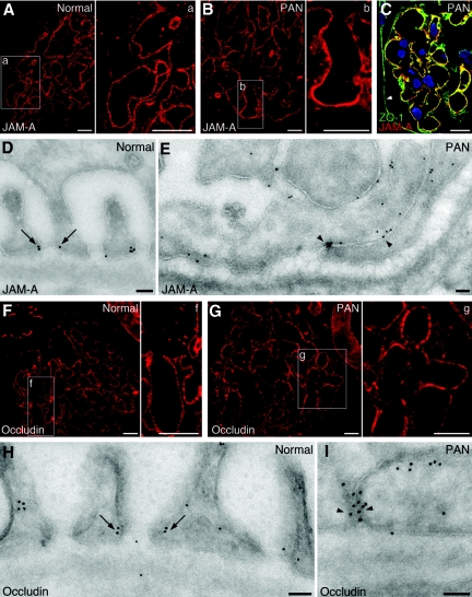
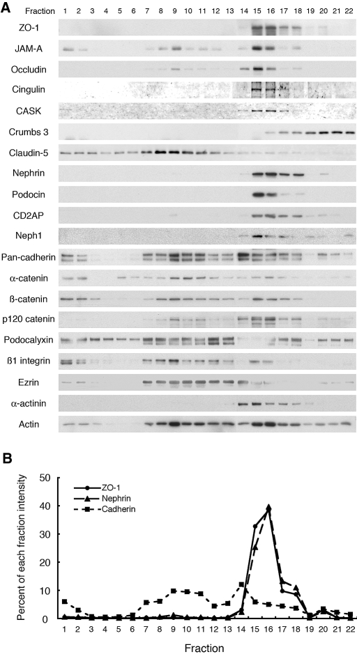
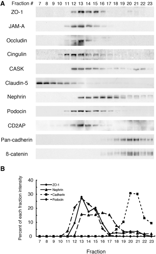
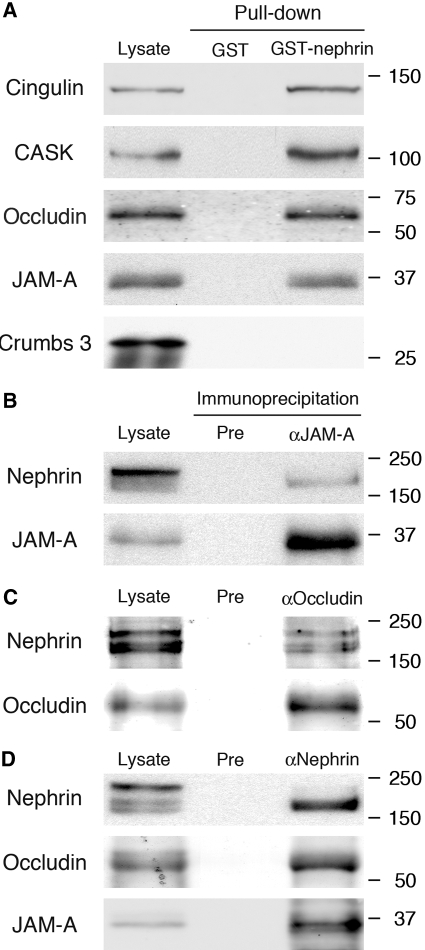
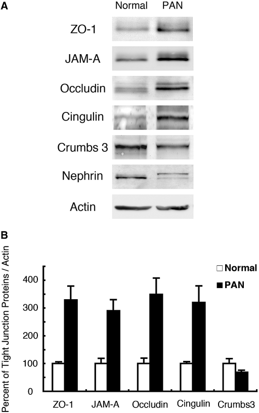
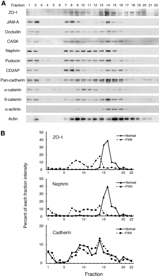
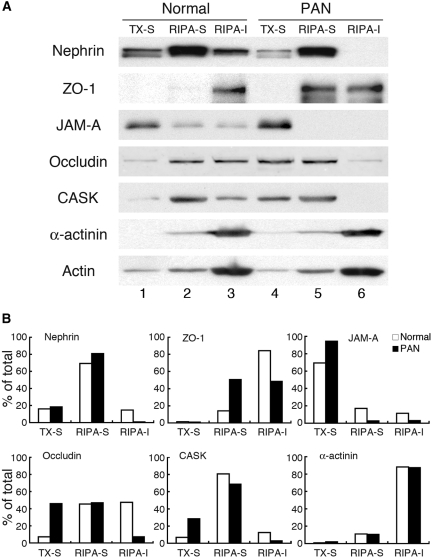
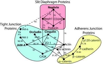
Slit diaphragms are essential components of the glomerular filtration apparatus, as changes in these junctions are the hallmark of proteinuric diseases. Slit diaphragms, considered specialized adherens junctions, contain both unique membrane proteins (e.g., nephrin, podocin, and Neph1) and typical adherens junction proteins (e.g., P-cadherin, FAT, and catenins). Whether slit diaphragms also contain tight junction proteins is unknown. Here, immunofluorescence, immunogold labeling, and cell fractionation demonstrated that rat slit diaphragms contain the tight junction proteins JAM-A (junctional adhesion molecule A), occludin, and cingulin. We found these proteins in the same protein complexes as nephrin, podocin, CD2AP, ZO-1, and Neph1 by cosedimentation, coimmunoprecipitation, and pull-down assays. PAN nephrosis increased the protein levels of JAM-A, occludin, cingulin, and ZO-1 several-fold in glomeruli and loosened their attachment to the actin cytoskeleton. These data extend current information about the molecular composition of slit diaphragms by demonstrating the presence of tight junction proteins, although slit diaphragms lack the characteristic morphologic features of tight junctions. The contribution of these proteins to the assembly of slit diaphragms and potential signaling cascades requires further investigation.


Figures

Figure 1.
Figure 1.
AM-A and occludin are present between foot processes of podocytes of normal and PAN nephrotic rats. By immunofluorescence, both JAM-A (A, a, and B, b) and occludin (F, f and G, g) are distributed in a punctate pattern along normal rat glomerular capillaries and in glomeruli from 7 d PAN-nephrotic rats (PAN). The boxed areas in A, B, F, and G are enlarged in a, b, f, and g. (C) By double labeling, JAM-A (red) and ZO-1 (green) colocalize (yellow) along glomerular capillaries at the base of the podocytes. Note that parietal cells (arrowhead) stain for ZO-1 but not for JAM-A. Nuclei were stained with DAPI (blue). (D) JAM-A and occludin (H) (10 nm gold) are seen to be concentrated at the slit diaphragms (arrows) in normal glomeruli. In PAN glomeruli, JAM-A (E) and occludin (I) are concentrated along the extensive tight or occluding junctions (arrowheads) (Bar = 10 μm in A-C and F-G; Bar = 50 nm in D, E, H, and I)
Figure 2.
Figure 2.
Distribution of junctional fractions from normal rat glomeruli in iodixanol density gradient centrifugation. (A) A Postnuclear supernatant (PNS) prepared from glomeruli isolated from normal rats was mixed with an equal amount of iodixanol [final concentration 30% (wt/vol)] and overlaid with equal volumes of 20% and 10% iodixanol. After centrifugation at 350,000 × g for 3 h at 4°C, fractions were collected from the top, separated by SDS-PAGE, and immunoblotted for the indicated 20 proteins expressed in foot processes of podocytes. In normal rat glomeruli, the tight junction proteins ZO-1, JAM-A, occludin, cingulin, and CASK cofractionate with the slit diaphragm enriched fractions marked by nephrin, podocin, CD2AP, and Neph1 in fractions 15 to 18. By contrast, the adherens junction proteins, cadherin and α-, β-, and p120 catenins, are broadly distributed in the iodixanol gradients (fractions 7 to 18) and only partially cofractionate with slit diaphragm proteins. Crumbs 3, another tight junction protein, is concentrated in heavier fractions (18–22) than other tight junction and slit diaphragm markers. Claudin-5, a specific marker for endothelial tight junctions, is concentrated in lighter fractions (1–12). Podocalyxin, an apical domain protein in foot processes, and β1 integrin, a basal protein, are broadly distributed in the gradient but are concentrated mostly in lighter fractions (1–13). Podocalyxin cofractionates with its binding partner, ezrin (8–14). (B) Densitometric analysis showing the % of the total nephrin (triangles with dashed line), pan-cadherin (squares with dotted line), and ZO-1 (circles with solid line) present in the fractions shown in “A.” The distribution of ZO-1 (used as a tight junction marker) in each of the fractions is similar to that of nephrin (slit diaphragm marker) but not to that of pan-cadherin (adherens junction marker).
Figure 3.
Figure 3.
Tight junction proteins cosediment in the same protein complexes as slit diaphragm proteins, but not adherens junction proteins. Glomeruli isolated from normal rats were lysed in 0.5% Nonidet P-40 and 0.25% Triton X-100, and the lysate was loaded on top of a 5 to 25% discontinuous sucrose gradient and centrifuged as described in Concise Methods. Fractions were collected from the top of the gradient and separated by SDS-PAGE followed by immunoblotting with the indicated antibodies. (A) ZO-1, JAM-A, occludin, and cingulin are concentrated in fractions 12 to 16 and cosediment with nephrin, podocin, CD2AP, and CASK. In contrast, cadherin and β-catenin distributed in heavier fractions (19–23) of the gradient. Claudin-5, a marker for endothelial tight junctions, is concentrated in lighter fractions (7–10). (B) Densitometric analysis of ZO-1 (circles with solid line), nephrin (triangles with dashed line), pan-cadherin (squares with dotted line), and podocin (diamonds with hatched line). ZO-1 partially cosediments with nephrin and podocin but not with pan-cadherin.
Figure 4.
Figure 4.
Tight junction proteins form a protein complex with nephrin but not crumbs 3. (A) JAM-A, occludin, cingulin, and CASK are pulled down with GST-nephrin tail from normal glomerular lysates. In contrast, crumbs 3 was not pulled down. Isolated glomeruli from normal rats were lysed in 1% Nonidet P-40 with 100 μmol/L potassium iodide. Twenty micrograms nephrin cytoplasmic domain fused to GST (GST-nephrin) or GST alone (GST) were incubated with 400 μg glomerular lysate at 4°C for 16 h and immunoblotted with JAM-A, occludin, cingulin, and CASK antibodies. (B to D) Nephrin can be coimmunoprecipitated with anti-JAM-A (B) and anti-occludin (C) IgG. Similarly, JAM-A and occludin can be coimmunopreciptated with anti-nephrin IgG (D). Isolated glomeruli from normal rats were lysed in 1% Triton X-100 and 0.5% Nonidet P-40. Immunoprecipitation was performed on 500 μg glomerular lysate from normal rats with JAM-A (αJAM-A), occludin (αOccludin), nephrin (αNephrin), or preimmune (Pre) IgG. Glomerular lysates (25 μg) were included as controls.
Figure 5.
Figure 5.
Comparative immunoblot analysis of expression of tight junction proteins in glomeruli from normal versus PAN nephrotic rats. (A) The protein levels of ZO-1, JAM-A, occludin, and cingulin are dramatically increased in glomerular lysates from PAN-treated rats (PAN) compared with normal, whereas expression of crumbs 3 and nephrin are decreased. β-actin was used as a control for protein load. Glomeruli isolated from two rats were lysed in RIPA buffer; 20 μg glomerular lysate was immunoblotted with the indicated antibodies. We performed experiments three times with similar results. (B) Densitometric analyses of data in “A” showing expression of tight junction proteins relative to β-actin in normal (open bars) and PAN (closed bars) glomeruli. Data are mean ± SEM values.
Figure 6.
Figure 6.
Iodixanol density gradient centrifugation of postnuclear supernatant prepared from glomeruli of PAN nephrotic rats. (A) In glomeruli from PAN-treated rats, fractions containing ZO-1, JAM-A, occludin, and CASK and the slit diaphragm markers, nephrin, podocin, and CD2AP, float up to regions of lower density and are broadly distributed in fractions 7 to 16. In normal glomeruli, they are restricted to fraction 15 to 18 (compare with Figure 2). In contrast, the distribution of adherens junction proteins (cadherins, α- and β-catenins) is similar in normal and PAN rats. (B) Densitometric analysis demonstrating that the distribution of nephrin and ZO-1 is shifted to lighter fractions in PAN (dashed lines) compared with normal (solid lines) glomeruli (data from Figure 2A). There is little change in the distribution of cadherins.
Figure 7.
Figure 7.
Sequential detergent extraction of proteins from glomeruli of normal and PAN-treated rats. (A) Immunoblot of normal and PAN-treated glomeruli after sequential detergent extraction. In normal glomeruli, nephrin, JAM-A, occludin, and CASK distributed in all three fractions (lanes 1 to 3), whereas ZO-1 and α-actinin mainly distributed in the RIPA-insoluble (RIPA-I) (lane 3) fraction. PAN treatment resulted in a significant increase in the detergent solubility of nephrin, ZO-1, JAM-A, occludin, and CASK in that increased amounts of these proteins were found in the Triton-soluble (compare lanes 1 and 4) and RIPA-soluble fraction (compare lanes 2 and 5), and only ZO-1 was detected in the RIPA-insoluble fraction (lane 6). The distribution of α-actinin and actin in each fraction is similar in normal and PAN rats. Glomeruli isolated from normal and PAN-treated rats were sequentially solubilized in 0.5% Triton X-100 and RIPA lysis buffer and separated into Triton X-100-soluble (TX-S), RIPA-soluble (RIPA-S), and RIPA-insoluble fractions, followed by immunoblotting. (B) Densitometric analysis showing the % of total protein in the Triton X-100-soluble, RIPA-soluble, and RIPA-insoluble fractions in normal (open bars) and PAN rats (closed bars) in “A.” Each bar indicates the percent of the total band intensity in each fraction.
Figure 8.
Figure 8.
Proposed protein-protein interaction map for the podoctye slit diaphragm. Interactions between proteins analyzed in this study are shown based on interactions listed in the I2D database, and validated in the literature (references shown). The slit diaphragm (red) and tight junction (blue) groups of proteins are linked by a direct interaction between Neph1 and ZO-1. This is the shortest link between nephrin and occludin, cingulin, and JAM-A (shown in bold). An alternative interaction may occur between nephrin and JAM-A via PAR-3 and PAR-6 (shown with dashed line and smaller font), which were not examined experimentally in the current study. In contrast to tight junction proteins, the adherens junction proteins studied lack direct interactions with slit diaphragm proteins, but indirect interactions are present (e.g., α-catenin is linked indirectly to CD2AP through actin and indirectly to Neph1 through ZO-1).

Comment in

References

    1. Pavenstadt H, Kriz W, Kretzler M: Cell biology of the glomerular podocyte. Physiol Rev 83: 253–307, 2003 - PubMed
    1. Reeves W, Caulfield JP, Farquhar MG: Differentiation of epithelial foot processes and filtration slits: Sequential appearance of occluding junctions, epithelial polyanion, and slit membranes in developing glomeruli. Lab Invest 39: 90–100, 1978 - PubMed
    1. Reeves WH, Kanwar YS, Farquhar MG: Assembly of the glomerular filtration surface. Differentiation of anionic sites in glomerular capillaries of newborn rat kidney. J Cell Biol 85: 735–753, 1980 - PMC - PubMed
    1. Caulfield JP, Reid JJ, Farquhar MG: Alterations of the glomerular epithelium in acute aminonucleoside nephrosis. Evidence for formation of occluding junctions and epithelial cell detachment. Lab Invest 34: 43–59, 1976 - PubMed
    1. Kurihara H, Anderson JM, Farquhar MG: Increased Tyr phosphorylation of ZO-1 during modification of tight junctions between glomerular foot processes. Am J Physiol 268: F514–524, 1995 - PubMed

Publication types

MeSH terms

LinkOut - more resources