. 2009 Aug;19(4):203-10.
doi: 10.1097/CMR.0b013e32832ccd6e.

Nuclear PTEN levels and G2 progression in melanoma cells

Affiliations

Nuclear PTEN levels and G2 progression in melanoma cells

Abraham I Jacob et al. Melanoma Res. 2009 Aug.

Abstract

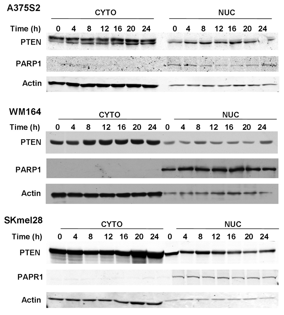
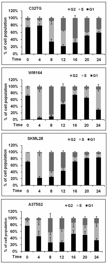
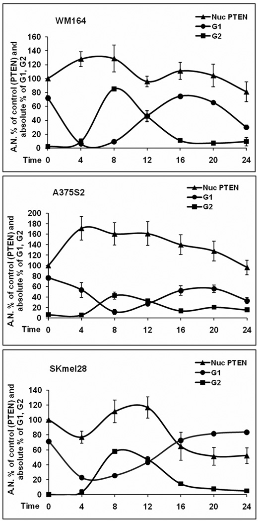
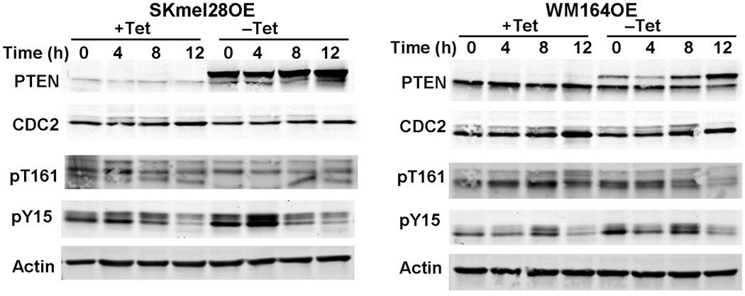
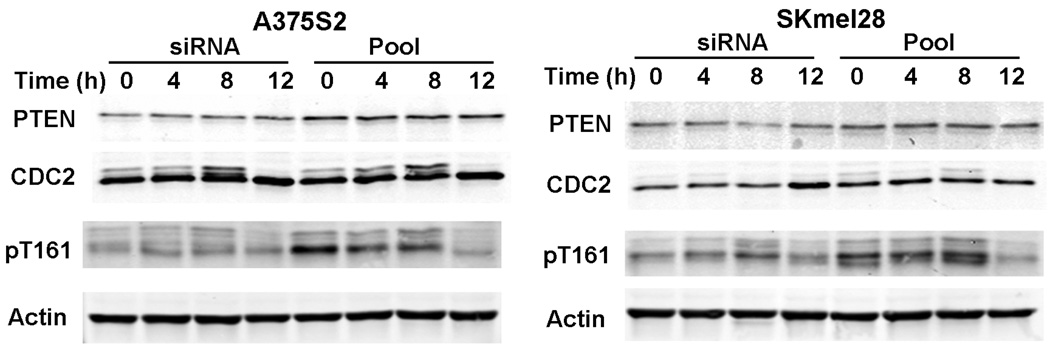
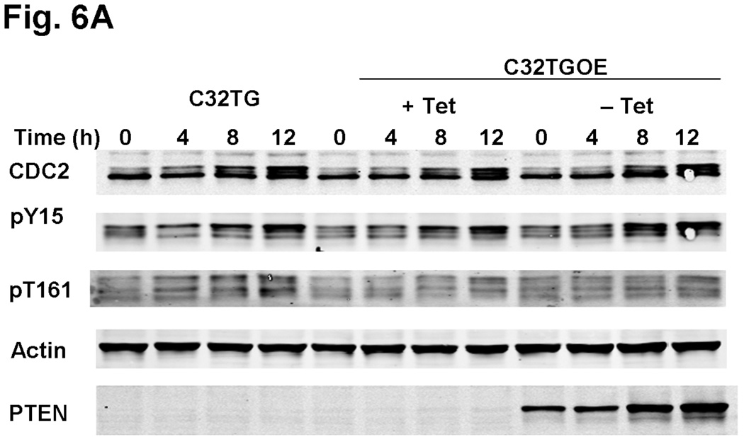
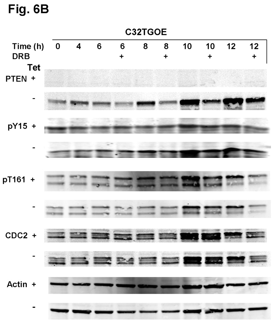
The phosphatase and tensin homolog (PTEN) exerts its function, in part, by negatively regulating the well-known phosphatidylinositol-3-kinase/AKT signaling pathway. Previous histological work has suggested that alterations in the nuclear/cytoplasmic compartmentalization of PTEN may play a role in the development and progression of melanoma. In this study, we examined the nuclear/cytoplasmic compartmentalization of PTEN in melanoma cell lines and its correlation with the cell cycle. Studies were performed in melanoma cells lines using classic cell biological techniques. In contrast to breast cancer cell lines, we found that increased levels of nuclear PTEN levels correlate with G2 rather than with G1 arrest. In WM164 and SKmel28 cells, overexpression of PTEN protein did not significantly increase the number of cells in the G2 phase. Differential CDC2 phosphorylation levels in cells that overexpressed PTEN compared with those where PTEN was downregulated suggest some involvement of PTEN in G2 checkpoint regulation. The data suggest that although nuclear PTEN levels correlate with the G2 phase, the role of PTEN in modulating G2/M arrest is not limiting. Further, the specific cell cycle phase regulated by nuclear PTEN is cell-type dependent. Taken together, our observations suggest that in melanoma, nuclear PTEN is involved in G2 progression possibly through the modulation of CDC2, opening up a new arena for investigation.


Conflict of interest statement

Conflict of interest: All of the authors declare no conflict of interest.

Figures

Fig. 1
Fig. 1
Nuclear (Nuc) and cytoplasmic (Cyto) PTEN levels change with cell cycle progression. Melanoma lines were grown to 60% confluence and synchronized with 3λmmol/l HU for 16–18λh. Cells were harvested at the time of release and at the indicated times, after release of HU. After subcellular fractionation, equal amounts of protein were separated on sodium dodecyl sulphate-polyacrylamide gel electrophoresis, transferred to membranes, and immunoblotted with specific anti-PTEN antibodies. Each blot is representative of five individual experiments. HU, hydroxyurea; PARP, poly (ADP-ribose) polymerase; PTEN, phosphatase and tensin homolog.
Fig. 2
Fig. 2
HU synchronized melanoma cells differ in G2 entrance. Cells of each melanoma cell line were grown to 60% confluence and treated with 3λmmol/l HU for 16–18λh. After HU release, cells were collected and analyzed by flow cytometry. Panels show the percent mean and SEM of cells in G1 (black bars, bottom), S (light gray bars), and G2 (dark gray bars, top) at each time point, for at least five individual experiments. HU, hydroxyurea.
Fig. 3
Fig. 3
Nuclear (Nuc) PTEN correlates with G2 population. After HU treatment and release, cells were subjected to subcellular fractionation and submitted to western blot or flow cytometry and quantitated. Shown are quantitated data from Figure 1, representing the nuclear fraction from 5–7 western blots, together with the cell cycle data from Figure 3, showing G1 and G2 from 5–7 experiments. For the western blots, time 0 was defined as control at 100%, and subsequent time points were calculated as percent of controls. Bars represent SEM and time in hours after HU release. HU, hydroxyurea.
Fig. 4
Fig. 4
Overexpression of PTEN decreases CDC2 phosphorylation. Cells overexpressing PTEN were grown to 60% confluence and synchronized with 3λmmol/l hydroxyurea for 16–18λh. After release and at each time point, cells were harvested and equal amounts of protein were separated by sodium dodecyl sulphate-polyacrylamide gel electrophoresis, transferred to membranes, and immunoblotted with the indicated antibodies. The blots in the figure are representative of five individual experiments. PTEN, phosphatase and tensin homolog; Tet, tetracycline; Skmel28OE, PTEN-overexpressing SKmel28; WM164OE, PTEN-overexpressing WM164 cells.
Fig. 5
Fig. 5
Downregulation of phosphatase and tensin homolog (PTEN) does not affect cell cycle. Cells treated with small interfering RNA (siRNA) were treated grown to 60% confluence and synchronized with 3λmmol/l hydroxyurea for 16–18λh. After release and at each time point, cells were harvested and equal amounts of protein were separated by sodium dodecyl sulphate-polyacrylamide gel electrophoresis, transferred to membranes, and immunoblotted with the indicated antibodies. The blots in this figure are representative of five individual experiments.
Fig. 6
Fig. 6
PTEN overexpressing C32TG cells show increased CDC2 tyrosine-15 phosphorylation. (a) C32TG cells overexpressing PTEN were grown to 60% confluence and synchronized with 3λmmol/l HU for 16–18λh. After release and at each time point, cells were harvested and equal amounts of protein were separated by sodium dodecyl sulphate-polyacrylamide gel electrophoresis, transferred to membranes, and immunoblotted with the indicated antibodies. Figure is representative of five individual experiments. (b) Cells were treated as in (a) with the following modification: starting 4λh after HU release, indicated cells (+) were treated with DRB until cells were harvested. Arrows indicate changes in phosphorylation status due to DRB treatment. C32TGOE, PTEN-expressing C32TG cells; DRB, 5,6-dichlorobenzimidazole riboside; HU, hydroxyurea; PTEN, phosphatase and tensin homolog; Tet, tetracycline.
Fig. 6
Fig. 6
PTEN overexpressing C32TG cells show increased CDC2 tyrosine-15 phosphorylation. (a) C32TG cells overexpressing PTEN were grown to 60% confluence and synchronized with 3λmmol/l HU for 16–18λh. After release and at each time point, cells were harvested and equal amounts of protein were separated by sodium dodecyl sulphate-polyacrylamide gel electrophoresis, transferred to membranes, and immunoblotted with the indicated antibodies. Figure is representative of five individual experiments. (b) Cells were treated as in (a) with the following modification: starting 4λh after HU release, indicated cells (+) were treated with DRB until cells were harvested. Arrows indicate changes in phosphorylation status due to DRB treatment. C32TGOE, PTEN-expressing C32TG cells; DRB, 5,6-dichlorobenzimidazole riboside; HU, hydroxyurea; PTEN, phosphatase and tensin homolog; Tet, tetracycline.

References

    1. Eng C. PTEN: one gene, many syndromes. Hum Mutat. 2003;22:183–198. - PubMed
    1. Liaw D, Marsh DJ, Li J, Dahia PL, Wang SI, Zheng Z, et al. Germline mutations of the PTEN gene in Cowden disease, an inherited breast and thyroid cancer syndrome. Nat Genet. 1997;16:64–67. - PubMed
    1. Marsh DJ, Dahia PL, Zheng Z, Liaw D, Parsons R, Gorlin RJ, et al. Germline mutations in PTEN are present in Bannayan-Zonana syndrome. Nat Genet. 1997;16:333–334. - PubMed
    1. Eng C, Thiele H, Zhou XP, Gorlin RJ, Hennekam RC, Winter RM. PTEN mutations and proteus syndrome. Lancet. 2001;358:2079–2080. - PubMed
    1. Zhou XP, Marsh DJ, Hampel H, Mulliken JB, Gimm O, Eng C. Germline and germline mosaic PTEN mutations associated with a Proteus-like syndrome of hemihypertrophy, lower limb asymmetry, arteriovenous malformations and lipomatosis. Hum Mol Genet. 2000;9:765–768. - PubMed

Publication types