Review
. 2009 Jun;6(6):415-22.
doi: 10.1038/nmeth.1332.

Transposon-mediated genome manipulation in vertebrates

Affiliations
Review

Transposon-mediated genome manipulation in vertebrates

Zoltán Ivics et al. Nat Methods. 2009 Jun.

Abstract

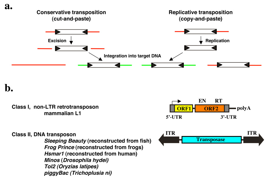
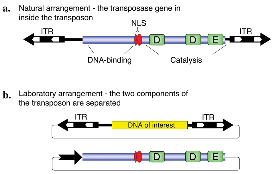
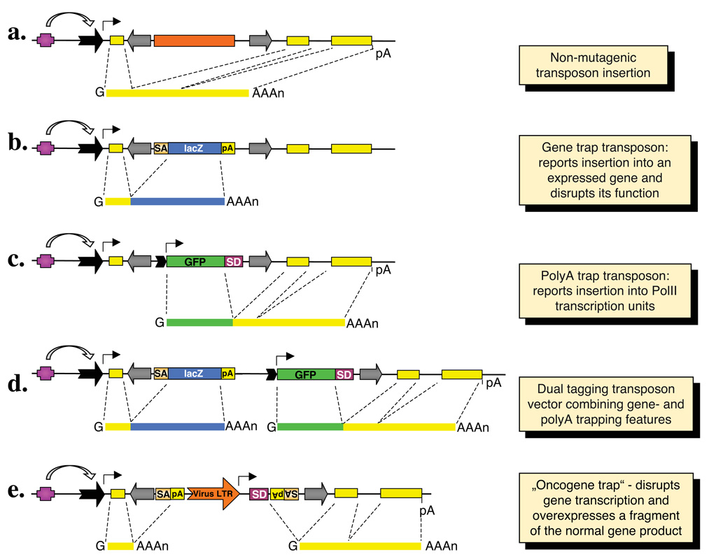
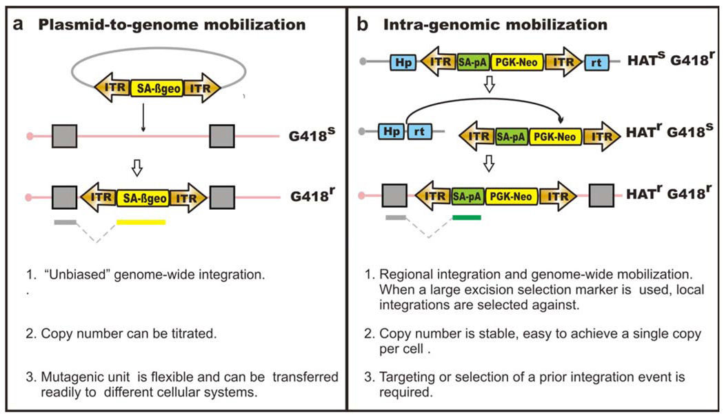
Transposable elements are DNA segments with the unique ability to move about in the genome. This inherent feature can be exploited to harness these elements as gene vectors for genome manipulation. Transposon-based genetic strategies have been established in vertebrate species over the last decade, and current progress in this field suggests that transposable elements will serve as indispensable tools. In particular, transposons can be applied as vectors for somatic and germline transgenesis, and as insertional mutagens in both loss-of-function and gain-of-function forward mutagenesis screens. In addition, transposons will gain importance in future cell-based clinical applications, including nonviral gene transfer into stem cells and the rapidly developing field of induced pluripotent stem cells. Here we provide an overview of transposon-based methods used in vertebrate model organisms with an emphasis on the mouse system and highlight the most important considerations concerning genetic applications of the transposon systems.


Figures

Figure 1
Figure 1. Mechanism of transposition and general organization of Class I and Class II transposable elements
(a) Schematic representation of the two major mechanisms of transposition. During conservative transposition, the element is excised from the donor DNA (red line), and integrates into a new target DNA (green line). Ligation of the broken ends of the DNA reconstitutes the donor locus. Replicative transposition involves amplification of the element by copying through transcription followed by reverse transcription. The newly made copy gets inserted elsewhere in the genome, but the donor element does not move. (b) Structures and organization of the main types of transposable elements. Class I non-LTR retrotransposon. The element consists of a 5’ untranslated region that has promoter activity (arrow) that drives transcription of the element-encoded genes. ORF1 encodes a nucleic acid binding protein. ORF2 encodes an endonuclease (EN) and a reverse transcriptase (RT). The element has a polyA tail. Class II DNA transposon. The central transposase gene is flanked by terminal inverted repeats (IRs, shown as black arrows). The IRs contain the binding sites for the transposase and sequences that are required for transposase-mediated cleavage.
Figure 2
Figure 2. Class II DNA transposon system
(a) Structure of the transposon. The central transposase gene (blue box) is flanked by terminal inverted repeats (IR, black arrows) that contain binding sites for the transposase (white arrows). In case of Sleeping Beauty, the transposase consists of an N-terminal DNA-binding domain, a nuclear localization signal (NLS) and a catalytic domain characterized by the DDE signature. (b) Gene transfer vector system based on a Class II DNA transposon. The transposase coding region can be replaced by a gene of interest (yellow box) within the transposable element. In a typical two-component, binary gene transfer vector system, the transposon can be mobilized if a transposase source is provided in cells; for example, the transposase can be expressed from a separate plasmid vector.
Figure 3
Figure 3. Summary of the basic gene trapping strategies
On top, a hypothetical transcription unit is depicted with an upstream regulatory element (purple box), a promoter (black arrow), three exons (yellow boxes) and a polyadenylation signal (pA). Major classes of transposon-based trapping constructs and spliced transcripts are shown below. Transposon inverted repeats are indicated by gray arrows. (a) An intronic transposon insertion is typically not mutagenic, because the transposon is spliced out from the primary RNA transcript together with the targeted intron sequences. (b) Gene trapping cassettes contain a splice acceptor (SA) followed by a reporter gene and a pA. The reporter is only expressed when transcription starts from the promoter of an endogenous transcription unit. Thus, the expression of the reporter follows the expression pattern of the trapped gene. (c) Polyadenylation [poly(A)] traps contain a promoter followed by a reporter gene and a splice donor (SD) site, but they lack a pA signal. Therefore, reporter gene expression depends on splicing to downstream exon/s of a Pol II transcription unit containing a pA. (d) The “dual tagging” vectors are based on both gene- and poly(A) trapping of a targeted transcription unit. (e) The oncogene trap contains SA signals followed by pA signals in both orientations to disrupt transcription, as well as a strong, viral enhancer/promoter that drives transcription towards the outside of an inserted transposon, and thereby overexpresses a gene product.
Figure 4
Figure 4. Transposon delivery methods in ES cells
Gene trap based loss-of-function mutagenesis is shown here as an example. (a) plasmid-to-genome mobilization. Cells with mutagenic transposon insertions can be selected in G418. (b) Intra-genomic mobilization. Upon transposase expression, the transposon will be excised from the donor site and re-integrate at a different genomic location. Enrichment of such cells can be achieved by selecting for transposition excision and reintegration. Using Hprt as an excision selection marker, cells with the transposon excised from the donor site will be Hprt-proficient and therefore HAT-resistant.
Figure 5
Figure 5. Recessive genetic screens in Blm-deficient ES cells
(a) Non-sister mitotic homologous recombination gives rise to homozygote mutants. The star represents the integration site of an intertional mutagen. The genotype highlighted in grey contains a bi-allelic mutation. (b) Flow chart for recessive genetic screens using a Blm-deficient ES cells system.
Figure 6
Figure 6. In vivo germline mutagenesis of the mouse with transposable elements
Breeding of “jumpstarter” and “mutator” stocks induces transposition in the germline of double-transgenic “seed” males. The transposition events that take place in germ cells are segregated in the offspring. Animals with transposition events need to be bred to homozygosity in order to visualize the phenotypic effects of recessive mutations. Mutant genes can easily be cloned by different PCR methods making use of the inserted transposon as a unique sequence tag.
Figure 7
Figure 7. Synthetic L1/ORFeus transgene and progeny retrotransposon insertions
(a) The transgene construct or donor element consists of the following sequence elements from 5’ to 3’: (i) a composite CMV IE enhancer/modified chicken β-actin promoter, designated “CAG”. (ii) synthetic L1 ORF1, ORF2 and 5’ portion of 3’UTR. (iii) Herpes simplex virus thymidine kinase poly(A) signal (boxed inverted letter A) in antisense orientation to polyadenylate gfp mRNA. (iv) gfp (green block arrow), a modified version of EGFP coding sequence. The gfp ORF is in antisense orientation relative to L1 and interrupted by intron 2 of human β-globin gene, which is in sense orientation relative to L1; gfp serves as a “retrotransposition indicator gene”. (v) Rous sarcoma virus LTR promoter in antisense orientation relative to L1, which drives gfp transcription (boxed inverted P for promoter). (vi) β-globin poly(A) signal (boxed upright letter A). Numbered arrows above the diagram represent locations of genotyping PCR primers. Region used to generate Southern blotting probes is indicated (purple line). (b) Structure of a representative progeny element. A typical progeny insertion is 5’ truncated, intronless, ends in a poly(A) tail (AAA) and is flanked by target site duplications (gray triangles) and target genomic DNA sequences (wavy solid lines). Primers 1 and 1’ (intron flanking primers) amplify a longer product when derived from the donor element (A) than from the progeny insertions (B); product length differs by the length of the intron. Primers 2 and 1’ (primer 2 is the “intron spanning” primer that spans the splice junction give rise to a product only from progeny retrotransposition events. Primers 3 and 3’ are control primers that give rise to products of constant length for donor and progeny elements.
Figure 8
Figure 8. Somatic mutagenesis in the mouse with transposable elements
Breeding of “jumpstarter” and “mutator” stocks induces transposition in the soma of double-transgenic animals (“oncomice”). In case of tissue-specific screens, a third genotype containing a tissue-specific Cre allele has to be crossed in. The crosses can be made either in wild-type or in specific cancer-predisposed genetic backgrounds. Transposition in somatic cells leads to random insertional mutations, and animals are aged for tumor development. Transposon insertions are cloned from genomic DNA isolated from tumor samples, and are subsequently mapped and annotated with respect to mutagenized genes. Those genes repeatedly mutated in multiple, independent tumors are designated as common insertion sites or CIS. These candidate cancer genes are functionally validated.

Similar articles

Cited by

References

    1. Lander ES, et al. Initial sequencing and analysis of the human genome. Nature. 2001;409:860–921. - PubMed
    1. Feng Q, Moran JV, Kazazian HH, Jr, Boeke JD. Human L1 retrotransposon encodes a conserved endonuclease required for retrotransposition. Cell. 1996;87:905–916. - PubMed
    1. Mathias SL, Scott AF, Kazazian HH, Jr, Boeke JD, Gabriel A. Reverse transcriptase encoded by a human transposable element. Science. 1991;254:1808–1810. - PubMed
    1. Luan DD, Korman MH, Jakubczak JL, Eickbush TH. Reverse transcription of R2Bm RNA is primed by a nick at the chromosomal target site: a mechanism for non-LTR retrotransposition. Cell. 1993;72:595–605. - PubMed
    1. Cost GJ, Feng Q, Jacquier A, Boeke JD. Human L1 element target-primed reverse transcription in vitro. Embo J. 2002;21:5899–5910. - PMC - PubMed

Substances