

Identification and functional distribution of intracellular ca channels in mouse lacrimal gland acinar cells
- PMID: 19478858
- PMCID: PMC2605693
- DOI: 10.2174/1874364100701010008
Identification and functional distribution of intracellular ca channels in mouse lacrimal gland acinar cells
Abstract
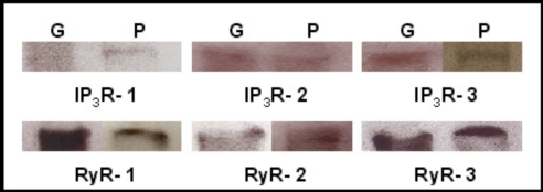
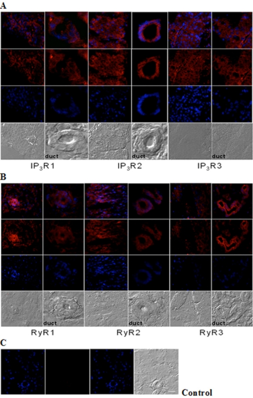
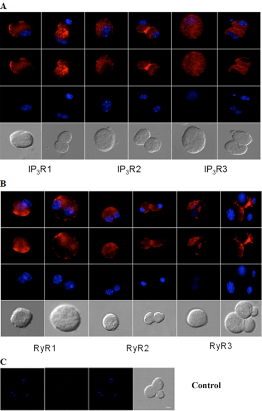
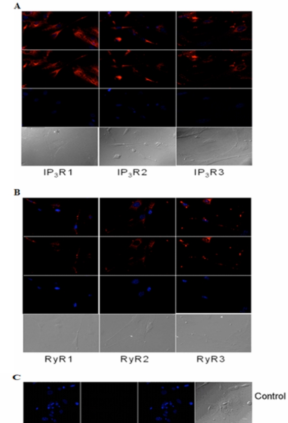
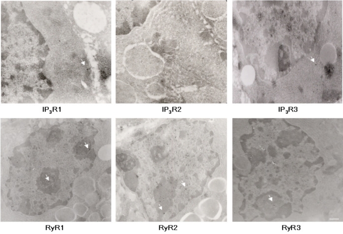
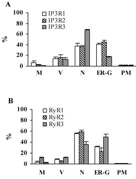
We have determined the presence and cellular distribution of intracellular calcium channels, inositol 1, 4, 5-trisphosphate receptors (IP3Rs) and ryanodine receptors (RyRs) in adult and postnatal (P10) lacrimal gland acinar cells. Western blot analysis of both P10 cultures and adult tissue identified the presence of each IP(3)R and RyR isotypes. The immunocytochemistry analysis showed a differential cellular distribution of these calcium channels where the nuclear envelope, endoplasmic reticulum (ER) and Golgi apparatus membranes represent areas with highest levels of channel expression. This IP(3)R and RyR isotype distribution is confirmed by the immuno-EM results. The findings described in this study are in agreement with published pharmacological data that shows the participation of these channels in the secretion process of the lacrimal gland acinar cells. Furthermore, the differential subcellular distribution between the isoforms could indicate a potential role of these intracellular Ca(2+ )channels on the regulation of specific cellular functions.
Figures
Similar articles
-
Polycystin-2 expression and function in adult mouse lacrimal acinar cells.Invest Ophthalmol Vis Sci. 2011 Jul 29;52(8):5605-11. doi: 10.1167/iovs.10-7114. Invest Ophthalmol Vis Sci. 2011. PMID: 21508103 Free PMC article.
-
Structural insights into endoplasmic reticulum stored calcium regulation by inositol 1,4,5-trisphosphate and ryanodine receptors.Biochim Biophys Acta. 2015 Sep;1853(9):1980-91. doi: 10.1016/j.bbamcr.2014.11.023. Epub 2014 Nov 25. Biochim Biophys Acta. 2015. PMID: 25461839 Review.
-
Novel biochemical and functional insights into nuclear Ca(2+) transport through IP(3)Rs and RyRs in osteoblasts.Am J Physiol Renal Physiol. 2000 May;278(5):F784-91. doi: 10.1152/ajprenal.2000.278.5.F784. Am J Physiol Renal Physiol. 2000. PMID: 10807590
-
Dynamic imaging of endoplasmic reticulum Ca2+ concentration in insulin-secreting MIN6 Cells using recombinant targeted cameleons: roles of sarco(endo)plasmic reticulum Ca2+-ATPase (SERCA)-2 and ryanodine receptors.Diabetes. 2002 Feb;51 Suppl 1:S190-201. doi: 10.2337/diabetes.51.2007.s190. Diabetes. 2002. PMID: 11815480
-
Sarco-Endoplasmic Reticulum Calcium Release Model Based on Changes in the Luminal Calcium Content.Adv Exp Med Biol. 2020;1131:337-370. doi: 10.1007/978-3-030-12457-1_14. Adv Exp Med Biol. 2020. PMID: 31646517 Review.
Cited by
-
Localization and phenotype-specific expression of ryanodine calcium release channels in C57BL6 and DBA/2J mouse strains.Exp Eye Res. 2011 Nov;93(5):700-9. doi: 10.1016/j.exer.2011.09.001. Epub 2011 Sep 14. Exp Eye Res. 2011. PMID: 21933672 Free PMC article.
-
P2Y purinoceptors induce changes in intracellular calcium in acinar cells of rat lacrimal glands.Histochem Cell Biol. 2012 Jan;137(1):97-106. doi: 10.1007/s00418-011-0885-0. Epub 2011 Nov 8. Histochem Cell Biol. 2012. PMID: 22065011
-
Ion channels in dry eye disease.Indian J Ophthalmol. 2023 Apr;71(4):1215-1226. doi: 10.4103/IJO.IJO_3020_22. Indian J Ophthalmol. 2023. PMID: 37026252 Free PMC article. Review.
-
Polycystin-2 expression and function in adult mouse lacrimal acinar cells.Invest Ophthalmol Vis Sci. 2011 Jul 29;52(8):5605-11. doi: 10.1167/iovs.10-7114. Invest Ophthalmol Vis Sci. 2011. PMID: 21508103 Free PMC article.
-
Calcium signaling in lacrimal glands.Cell Calcium. 2014 Jun;55(6):290-6. doi: 10.1016/j.ceca.2014.01.001. Epub 2014 Jan 22. Cell Calcium. 2014. PMID: 24507443 Free PMC article. Review.
References
-
- Hodges RR, Dartt DA. Regulatory pathways in lacrimal gland epithelium. Int Rev Cytol. 2003;231:129–96. - PubMed
-
- Paranyuk Y, Claros N, Birzgalis A, Moore LC, Brink PR, Walcott B. Lacrimal gland fluid secretion and lymphocytic infiltration in the NZB/W mouse model of Sjögren's syndrome. Curr Eye Res. 2001;23(3):199–205. - PubMed
-
- Walcott B. The lacrimal gland and its veil of tears. News Physiol Sci. 1998;13:97–103. - PubMed
-
- Bromberg BB. Autonomic control of lacrimal protein secretion. IOVS. 1981;20:110–116. - PubMed
-
- Sundermeier T, Matthews G, Brink PR, Walcott B. Calcium dependence of exocytosis in lacrimal gland acinar cells. Am J Physiol Cell Physiol. 2002;282:C360–C365. - PubMed
Grants and funding
LinkOut - more resources
Full Text Sources
Miscellaneous