

Histological analysis of the lower-positioned transverse ligament
- PMID: 19478863
- PMCID: PMC2605698
- DOI: 10.2174/1874364100701010017
Histological analysis of the lower-positioned transverse ligament
Abstract
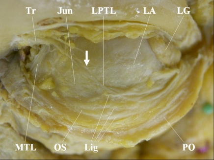
The lower-positioned transverse ligament (LPTL) had been thought to run parallel to the junction between the orbital septum and the levator aponeurosis (junction). However, its true course was disclosed as crossing the junction. Since earlier histological studies were undertaken before the precise course was elucidated, it was uncertain whether the true LPTL was adequately disclosed. Therefore, we examined ten upper eyelids of 6 Asian patients who underwent blepharoptosis repairs. The LPTL and the tissue running parallel to the junction were harvested intraoperatively. Light-microscopically, the LPTL contained looser and thinner collagen bundles and less elastic fibres than the parallel tissue. Electron-microscopically, collagen microfibrils in the LPTL had almost the same periodicity and thickness as those in the parallel tissue. The LPTL is a loose and inelastic structure, which at a light microscopic level is completely different from the parallel tissue; however, the differences could not be verified by electron microscopy.
Figures


Similar articles
-
Aesthetic blepharoptosis correction with release of fibrous web bands between the levator aponeurosis and orbital fat.J Craniofac Surg. 2012 Jan;23(1):e52-5. doi: 10.1097/SCS.0b013e3182418d1a. J Craniofac Surg. 2012. PMID: 22337465
-
The levator aponeurosis consists of two layers that include smooth muscle.Ophthalmic Plast Reconstr Surg. 2005 Sep;21(5):379-82. Ophthalmic Plast Reconstr Surg. 2005. PMID: 16234705
-
Ultrasound biomicroscopy of the levator aponeurosis in congenital and aponeurotic blepharoptosis.Ophthalmic Plast Reconstr Surg. 2004 Jul;20(4):308-11. doi: 10.1097/01.iop.0000129532.33913.e7. Ophthalmic Plast Reconstr Surg. 2004. PMID: 15266146
-
[The three-dimensional ultrastructure of the collagen fibers, reticular fibers and elastic fibers: a review].Kaibogaku Zasshi. 1992 Jun;67(3):186-99. Kaibogaku Zasshi. 1992. PMID: 1523957 Review. Japanese.
-
Structural biology of the fibres of the collagenous and elastic systems.Cell Biol Int. 1996 Jan;20(1):15-27. doi: 10.1006/cbir.1996.0004. Cell Biol Int. 1996. PMID: 8936403 Review.
References
-
- Kakizaki H, Zako M, Miyaishi O, et al. Posterior aspect of the orbital septum is reinforced by ligaments. Jpn J Ophthalmol. 2005;49:477–80. - PubMed
-
- Kakizaki H, Zako M, Nakano T, et al. Modified course of the lower-positioned transverse ligament. Br J Plast Surg. 2005;58:1035–6. - PubMed
-
- Yuzuriha S, Matsuo K, Kushima H. An anatomical structure, which results in puffiness of the upper eyelid and a narrow palpebral fissure in the Mongoloid eye. Br J Plast Surg. 2000;53:466–72. - PubMed
-
- Anderson RL, Dixon RS. The role of Whitnall's ligament in ptosis surgery. Arch Ophthalmol. 1979;97:705–7. - PubMed
-
- Ettl A, Zonneveld F, Daxer A, Koornneef L. Is Whitnall's ligament responsible for the curved course of the levator palpebral superioris muscle? Ophthalmic Res. 1998;30:321–6. - PubMed
LinkOut - more resources
Full Text Sources