

The cortical topography of visual evoked potentials elicited by chromatic and luminance motion
- PMID: 19478864
- PMCID: PMC2605699
- DOI: 10.2174/1874364100701010025
The cortical topography of visual evoked potentials elicited by chromatic and luminance motion
Abstract
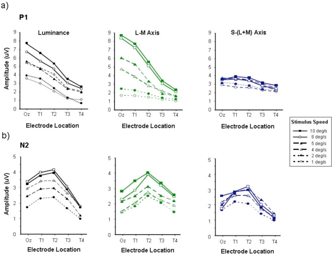
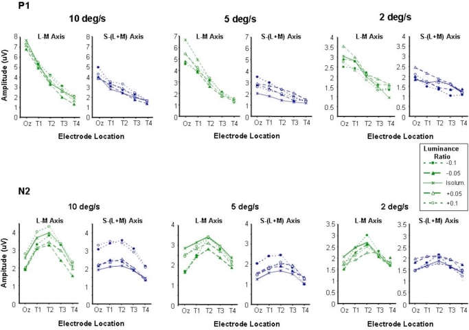
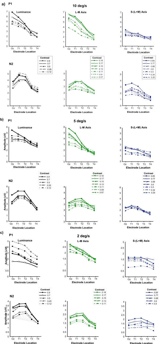
When motion-onset VEPs are elicited by moving luminance patterns, the motion specific component of the response, N2, is more prominent at electrode sites that overlay the lateral occipito-parietal cortex close to area V5/MT, than over the primary visual cortex. Functional segregation suggests that colour and motion processing should take place along different ventral occipito-temporal and lateral occipito-parietal pathways, respectively. Hence, a different topographical distribution might be expected for the motion-onset VEPs elicited by chromatic and luminance motion stimuli. We recorded motion-onset VEPs to moving luminance or isoluminant chromatic sinusoidal grating stimuli from five electrodes sites located at Oz, and at four locations (T1-T4) lateral to Oz, at intervals of 5% of the head circumference. Responses were recorded from 6 subjects over a range of speeds and contrasts. The results showed that the N2 component was maximal at similar lateral electrode locations (T2) for both luminance-defined and chromatically-defined motion. The earlier P1 component was of greatest magnitude at the occipital pole (Oz) and decreased with more lateral electrode placement and again this was the same for colour and luminance responses. These similarities suggest a common origin for VEPs elicited by colour and luminance defined motion.
Keywords: Colour; motion; visual evoked potentials (VEPs)..
Figures

Similar articles
-
Motion adaptation in chromatic motion-onset visual evoked potentials.Doc Ophthalmol. 2001 Nov;103(3):229-51. doi: 10.1023/a:1013037101814. Doc Ophthalmol. 2001. PMID: 11824660
-
Visual evoked potentials elicited by chromatic motion onset.Vision Res. 2001 Jul;41(15):2005-25. doi: 10.1016/s0042-6989(01)00080-3. Vision Res. 2001. PMID: 11412889
-
Development of the temporal properties of visual evoked potentials to luminance and colour contrast in infants.Vision Res. 1996 Oct;36(19):3141-55. doi: 10.1016/0042-6989(96)00050-8. Vision Res. 1996. PMID: 8917775
-
Visual evoked potentials in three-dimensional color space: correlates of spatio-chromatic processing.Vision Res. 1994 Oct;34(20):2657-71. doi: 10.1016/0042-6989(94)90222-4. Vision Res. 1994. PMID: 7975303
-
Motion-onset VEPs: characteristics, methods, and diagnostic use.Vision Res. 2007 Jan;47(2):189-202. doi: 10.1016/j.visres.2006.09.020. Epub 2006 Nov 28. Vision Res. 2007. PMID: 17129593 Review.
References
-
- Müller R, Göpfert E. The influence of grating contrast on the human cortical potential visually evoked by motion. Acta Neurobiol Exp. 1988;48:239–249. - PubMed
-
- Müller R, Göpfert E, Schlykowa L, Anke D. The human motion VEP as a function of size and eccentricity of the stimulation field. Doc Ophthalmol. 1990;76:81–89. - PubMed
-
- Göpfert E, Müller R, Simon EM. The human motion onset VEP as a function of stimulation area for foveal and peripheral vision. Doc Ophthalmol. 1990;75:165–173. - PubMed
-
- Kuba M, Kubova Z. Visual evoked potentials specific for motion onset. Doc Ophthalmol. 1992;80:83–89. - PubMed
-
- Bach M, Ullrich D. Motion adaptation governs the shape of motion evoked cortical potentials. Vis Res. 1992;34:1541–1547. - PubMed
LinkOut - more resources
Full Text Sources