. 2009 May;5(5):e1000450.
doi: 10.1371/journal.ppat.1000450. Epub 2009 May 29.

Vpu antagonizes BST-2-mediated restriction of HIV-1 release via beta-TrCP and endo-lysosomal trafficking

Affiliations

Vpu antagonizes BST-2-mediated restriction of HIV-1 release via beta-TrCP and endo-lysosomal trafficking

Richard S Mitchell et al. PLoS Pathog. 2009 May.

Abstract

The interferon-induced transmembrane protein BST-2/CD317 (tetherin) restricts the release of diverse enveloped viruses from infected cells. The HIV-1 accessory protein Vpu antagonizes this restriction by an unknown mechanism that likely involves the down-regulation of BST-2 from the cell surface. Here, we show that the optimal removal of BST-2 from the plasma membrane by Vpu requires the cellular protein beta-TrCP, a substrate adaptor for a multi-subunit SCF E3 ubiquitin ligase complex and a known Vpu-interacting protein. beta-TrCP is also required for the optimal enhancement of virion-release by Vpu. Mutations in the DSGxxS beta-TrCP binding-motif of Vpu impair both the down-regulation of BST-2 and the enhancement of virion-release. Such mutations also confer dominant-negative activity, consistent with a model in which Vpu links BST-2 to beta-TrCP. Optimal down-regulation of BST-2 from the cell surface by Vpu also requires the endocytic clathrin adaptor AP-2, although the rate of endocytosis is not increased; these data suggest that Vpu induces post-endocytic membrane trafficking events whose net effect is the removal of BST-2 from the cell surface. In addition to its marked effect on cell-surface levels, Vpu modestly decreases the total cellular levels of BST-2. The decreases in cell-surface and intracellular BST-2 are inhibited by bafilomycin A1, an inhibitor of endosomal acidification; these data suggest that Vpu induces late endosomal targeting and partial degradation of BST-2 in lysosomes. The Vpu-mediated decrease in surface expression is associated with reduced co-localization of BST-2 and the virion protein Gag along the plasma membrane. Together, the data support a model in which Vpu co-opts the beta-TrCP/SCF E3 ubiquitin ligase complex to induce endosomal trafficking events that remove BST-2 from its site of action as a virion-tethering factor.


Conflict of interest statement

The authors have declared that no competing interests exist.

Figures

Figure 1
Figure 1. β-TrCP is required for the optimal down-regulation of cell-surface BST-2: inhibition of Vpu activity by ΔF-box β-TrCP and shRNA targeting β-TrCP-1 and -2.
(A) ΔF-box β-TrCP inhibits Vpu-mediated down-regulation of cell-surface BST-2. Cells (HeLa) were transfected with either an empty plasmid or a plasmid expressing Vpu, along with a plasmid expressing GFP as a transfection marker. The cells were also transfected with either an empty plasmid (“mock”), a plasmid expressing β-TrCP, or a plasmid expressing a β-TrCP protein lacking the F-box (ΔFbox β-TrCP). The next day, the cells were stained for surface BST-2 and analyzed by two-color flow cytometry. Histograms represent the relative cell number vs. BST-2 fluorescence intensity for the GFP-positive cells. The percentage of GFP-positive cells varied between 30 and 33% for the six transfections shown. Gray-shaded histograms represent cells not transfected to express Vpu; unshaded histograms represent cells transfected to express Vpu. ΔF-box β-TrCP inhibited the Vpu-mediated down-regulation of cell surface BST-2 in each of four experiments; statistical analysis is described in the text. (B) Cells (HeLa) were transfected with a plasmid expressing β-TrCP (WT β-TrCP), or a plasmid expressing a β-TrCP protein lacking the F-box (ΔFbox β-TrCP), or not transfected; cell lysates were analyzed by immunoblot to detect the HA-tagged β-TrCP proteins. (C) shRNA targeting β-TrCP inhibits Vpu-mediated down-regulation of cell-surface BST-2. Cells (HeLa) were transfected with plasmids expressing shRNAs targeting either Renilla GFP (rGFP) as an irrelevant control, β-TrCP-1, β-TrCP-2, or both β-TrCP-1 and -2; in two cases these plasmids also expressed jellyfish GFP (the plasmids targeting β-TrCP-1 and both β-TrCP-1 and -2); for the others a separate plasmid expressing GFP was co-transfected. Two days later, the cells were re-transfected with an empty plasmid or a plasmid expressing Vpu, along with a plasmid expressing Tac antigen (IL-2 receptor α; CD25) as a transfection marker. The next day, the cells were stained for surface BST-2 and Tac, and then analyzed by three-color flow cytometry. Two-color dot plots are the BST-2 vs.Tac intensity of the individual GFP-positive cells. The results shown are representative of two independent experiments. (D) HeLa cells were transfected with the indicated plasmids expressing shRNAs used in (C) along with the plasmid expressing β-TrCP-1-HA; cell lysates were analyzed by immunoblot to detect the HA-tagged β-TrCP.
Figure 2
Figure 2. ΔF-box β-TrCP inhibits Vpu-mediated enhancement of virion-release.
Cells (HeLa) were transfected with either a proviral plasmid expressing wild-type HIV-1NL4-3 (“WT”) or a proviral plasmid expressing an isogenic vpu-negative mutant (“ΔVpu”). The cells were also transfected with an empty plasmid (“mock”), a plasmid expressing β-TrCP, or a plasmid expressing the β-TrCP ΔF-Box mutant. The next day, the fraction of the total p24 capsid antigen produced by the cells that was secreted into the media was measured. The average values from two independent experiments are graphed; the error bars indicate the actual values obtained from each experiment.
Figure 3
Figure 3. Residues within the DSGxxS β-TrCP binding motif of Vpu are required for optimal down-regulation of BST-2 and enhancement of virion-release.
(A) Down-regulation of BST-2 by Vpu-mutants. Cells (HeLa) were transfected with an empty plasmid, a plasmid expressing Vpu, or a plasmid expressing the indicated Vpu mutant, along with a plasmid expressing GFP as a transfection marker. The amount of Vpu-expression plasmid in each transfection (160 ng) was just sufficient in the case of the wild-type to rescue the release of virions from cells expressing a vpu-negative genome (see (C) below and the Materials and Methods section). The next day, the cells were stained for surface BST-2 and analyzed by two-color flow cytometry. Left: Histograms represent the relative cell number vs. BST-2 fluorescence intensity for the GFP-positive cells. In each panel, the heavy line is the curve for the indicated Vpu mutant. The shaded curve is the empty vector control, and the light line is the curve for wild-type Vpu. The percentage of cells that were GFP-positive was 11 for Vpu-WT, 14 for Vpu-D51A, 10 for Vpu-S52/56N, and 10 for Vpu-D51A-S52/56N. The results shown are representative of two independent experiments. Right: aliquots of each population were also analyzed by SDS-PAGE and immunoblot for Vpu and actin; molecular weight markers are indicated on the left in kilodaltons. (B) Vpu-S52/56N inhibits down-regulation of BST-2 by the wild-type protein. Cells (HeLa) were transfected as described in (A) above, except that in the right panel a combination of the plasmid expressing wild-type Vpu (160 ng) and the plasmid expressing Vpu-S52/56N (1.0 µg) was used (see also the Materials and Methods section). The next day, the cells were stained for surface BST-2 and analyzed by two-color flow cytometry. Histograms represent the relative cell number vs. BST-2 fluorescence intensity for the GFP-positive cells. The gray-shaded histogram represents cells not transfected to express Vpu (same in each panel); the unshaded histograms represent cells transfected to express Vpu, Vpu-S52/56N, or the combination of WT-Vpu plus Vpu-52/56N. The percentage of GFP-positive cells was 35 for WT-Vpu, 48 for Vpu-52/56N, and 49 for WT-Vpu plus Vpu-52/56N. The results shown are representative of two independent experiments. (C) Enhancement of virion-release by Vpu-mutants. Cells (HeLa) were transfected with a proviral plasmid expressing the vpu-negative mutant ΔVpu (1.44 µg), along with a plasmid expressing Vpu or the indicated Vpu mutant (160 ng). For the positive control, cells were transfected with the wild-type proviral plasmid alone; for the negative control, cells were transfected with ΔVpu along with an empty plasmid. The next day, the fraction of the total p24 capsid antigen produced by the cells that was secreted into the media was measured. Results are the average of duplicate transfections and are representative of two independent experiments.
Figure 4
Figure 4. Vpu decreases total cellular BST-2 to a lesser extent than cell-surface BST-2.
(A) Effect of Vpu on the steady-state total cellular levels of BST-2 detected by immunoblot. Cells (HeLa) were transfected with an empty plasmid or a plasmid expressing Vpu along with a plasmid expressing GFP as a transfection marker in a 20∶1 weight ratio. Left: The next day, the cells were stained for surface BST-2 and analyzed by two-color flow cytometry: two-color dot plots are the BST-2 vs. GFP intensity of the individual cells. Right: aliquots of each population were also analyzed by SDS-PAGE and immunoblot for Vpu, actin and for BST-2; molecular weight markers are indicated on the left in kilodaltons. (B) Effect of Vpu on intracellular and surface levels of BST-2 measured by flow cytometry. Cells (HeLa) were transfected as described in (A) with an empty plasmid or a plasmid expressing Vpu along with a plasmid expressing GFP as a transfection marker. The next day, the cells were stained for BST-2 either without (“surface”) or with (“intracellular”) permeabilization and analyzed by two-color flow cytometry: two-color plots are the BST-2 vs. GFP intensity of the individual cells.
Figure 5
Figure 5. The plasma membrane clathrin adaptor protein complex AP-2 is required for optimal down-regulation of BST-2 from the cell surface by Vpu.
(A) Cells (HeLa) were transfected once with siRNAs targeting either the medium (μ) subunits of AP-1 (μ1), AP-2 (μ2), AP-3 (μ3), or an irrelevant “non-target” (NT) sequence. Three days later, the cells were re-transfected with either an empty plasmid or a plasmid expressing Vpu, along with a plasmid expressing GFP as a transfection marker. The next day, the cells were stained for surface BST-2 and analyzed by two-color flow cytometry. Histograms represent the relative cell number vs. BST-2 fluorescence intensity for the GFP-positive cells. Gray-shaded histograms represent cells not transfected to express Vpu; unshaded histograms represent cells transfected to express Vpu. Inhibition of Vpu-mediated down-regulation of BST-2 by siRNA targeting μ2 was observed in each of four independent experiments. (B) Cells (HeLa) were transfected once with siRNAs targeting either μ2 or an irrelevant “non-target” (NT) sequence. Three days later, the cells were re-transfected with a plasmid expressing Vpu. The next day, the cells were fixed, permeabilized, and stained for Vpu and AP-2. The cells were imaged as described in the Materials and Methods section; a single focal plane is shown. Asterisks indicate cells with reduced expression of AP-2.
Figure 6
Figure 6. Vpu does not increase the rate of endocytosis of BST-2.
Cells (HeLa) were transfected with either an empty plasmid (“no Vpu”) or a plasmid expressing Vpu (“plus Vpu”), along with a plasmid expressing GFP as a transfection marker. The next day, the cells were labeled at 4°C with an antibody to BST-2, warmed for the indicated times at 37°C, then stained with a fluorophore-conjugated secondary antibody and analyzed by two-color flow cytometry. The amount of BST-2 remaining on the cell surface over time is shown for the GFP-positive cells. The fluorescence intensities of the time zero cells (no incubation at 37°C) for each population (“no Vpu” and “plus Vpu”) were set at 100%. The expression of Vpu reduced the surface levels of BST-2 by 3-fold in this experiment (data not shown). The results shown are representative of two independent experiments.
Figure 7
Figure 7. Bafilomycin A1 inhibits the ability of Vpu to down-regulate BST-2.
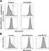
(A) Cells (HeLa) were transfected with either an empty plasmid or a plasmid expressing Vpu, along with a plasmid expressing GFP as a transfection marker. Immediately after the transfection, the cells were treated with bafilomycin A1 (final concentration 0.13 µM in DMSO) or DMSO only for 14 hours, and then stained for surface or intracellular BST-2 and analyzed by two-color flow cytometry. Histograms represent the relative cell number vs. BST-2 fluorescence intensity for the GFP-positive cells. Gray-shaded histograms represent cells transfected to express Vpu; unshaded histograms represent cells transfected with the empty plasmid. The plots shown are representative of at least two transfections for each experimental condition. (B) Cells (HeLa) were transfected as described above. Immediately after the transfection, the cells were treated with bafilomycin A1 (final concentration 0.13 µM in DMSO), MG-132 (final concentration 30 µM in DMSO), or DMSO only for 14 hours, and then stained for surface BST-2 and analyzed by two-color flow cytometry. Histograms represent the relative cell number vs. BST-2 fluorescence intensity for the GFP-positive cells. Gray-shaded histograms represent cells transfected to express Vpu; unshaded histograms represent cells transfected with the empty plasmid. The plots shown are representative of two independent experiments.
Figure 8
Figure 8. Vpu decreases the co-localization of BST-2 and the virion-protein p17 Gag along the plasma membrane.
(A) Cells (HeLa) were transfected to express either wild-type (vpu-positive) or vpu-negative (ΔVpu) viral genomes. The next day, the cells were fixed and stained without permeabilization for surface BST-2 (red). The cells were subsequently permeabilized with detergent and stained for p17/p55 Gag (blue). Images were acquired in a focal plane just above the cover glass to capture the distribution of proteins along the plasma membrane. (B) Correlation of the relative staining intensities of BST-2 and Gag in cell surface puncta; units are arbitrary.
Figure 9
Figure 9. Model for the relief of BST-2-mediated restriction by Vpu.
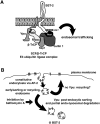
(A) Vpu recruits β-TrCP to induce ubiquitin-mediated trafficking events that remove BST-2 from the plasma membrane, its site of action as a virion-tethering factor. Circles in the cytoplasmic domain of Vpu represent phosphoserines 52 and 56. The interaction between BST-2 and Vpu and the ubiquitination of BST-2 are currently speculative. (B) Vpu induces bafilomycin A1-sensitive post-endocytic trafficking of BST-2 and endo-lysosomal degradation. The removal of BST-2 from the plasma membrane involves constitutive endocytosis of BST-2 via AP-2, followed by Vpu-mediated post-endocytic sorting events. Recycling of BST-2 to the plasma membrane in the absence of Vpu is currently speculative.

Similar articles

Cited by

References

    1. Malim MH, Emerman M. HIV-1 accessory proteins–ensuring viral survival in a hostile environment. Cell Host Microbe. 2008;3:388–398. - PubMed
    1. Yu X, Yu Y, Liu B, Luo K, Kong W, et al. Induction of APOBEC3G ubiquitination and degradation by an HIV-1 Vif-Cul5-SCF complex. Science. 2003;302:1056–1060. - PubMed
    1. Harris RS, Bishop KN, Sheehy AM, Craig HM, Petersen-Mahrt SK, et al. DNA deamination mediates innate immunity to retroviral infection. Cell. 2003;113:803–809. - PubMed
    1. Neil SJ, Zang T, Bieniasz PD. Tetherin inhibits retrovirus release and is antagonized by HIV-1 Vpu. Nature. 2008;451:425–430. - PubMed
    1. Van Damme N, Goff D, Katsura C, Jorgenson RL, Mitchell R, et al. The interferon-induced protein BST-2 restricts HIV-1 release and is downregulated from the cell surface by the viral Vpu protein. Cell Host Microbe. 2008;3:245–252. - PMC - PubMed

Publication types

MeSH terms

Substances