Cdc7p-Dbf4p regulates mitotic exit by inhibiting Polo kinase
- PMID: 19478884
- PMCID: PMC2682205
- DOI: 10.1371/journal.pgen.1000498
Cdc7p-Dbf4p regulates mitotic exit by inhibiting Polo kinase
Abstract
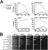
Cdc7p-Dbf4p is a conserved protein kinase required for the initiation of DNA replication. The Dbf4p regulatory subunit binds Cdc7p and is essential for Cdc7p kinase activation, however, the N-terminal third of Dbf4p is dispensable for its essential replication activities. Here, we define a short N-terminal Dbf4p region that targets Cdc7p-Dbf4p kinase to Cdc5p, the single Polo kinase in budding yeast that regulates mitotic progression and cytokinesis. Dbf4p mediates an interaction with the Polo substrate-binding domain to inhibit its essential role during mitosis. Although Dbf4p does not inhibit Polo kinase activity, it nonetheless inhibits Polo-mediated activation of the mitotic exit network (MEN), presumably by altering Polo substrate targeting. In addition, although dbf4 mutants defective for interaction with Polo transit S-phase normally, they aberrantly segregate chromosomes following nuclear misorientation. Therefore, Cdc7p-Dbf4p prevents inappropriate exit from mitosis by inhibiting Polo kinase and functions in the spindle position checkpoint.
Conflict of interest statement
The authors have declared that no competing interests exist.
Figures


Similar articles
-
Characterization of the yeast Cdc7p/Dbf4p complex purified from insect cells. Its protein kinase activity is regulated by Rad53p.J Biol Chem. 2000 Nov 10;275(45):35051-62. doi: 10.1074/jbc.M003491200. J Biol Chem. 2000. PMID: 10964916
-
A Dbf4p BRCA1 C-terminal-like domain required for the response to replication fork arrest in budding yeast.Genetics. 2006 Jun;173(2):541-55. doi: 10.1534/genetics.106.057521. Epub 2006 Mar 17. Genetics. 2006. PMID: 16547092 Free PMC article.
-
Cdc7p-Dbf4p kinase binds to chromatin during S phase and is regulated by both the APC and the RAD53 checkpoint pathway.EMBO J. 1999 Oct 1;18(19):5334-46. doi: 10.1093/emboj/18.19.5334. EMBO J. 1999. PMID: 10508166 Free PMC article.
-
A Cdc7p-Dbf4p protein kinase activity is conserved from yeast to humans.Prog Cell Cycle Res. 2000;4:61-9. doi: 10.1007/978-1-4615-4253-7_6. Prog Cell Cycle Res. 2000. PMID: 10740815 Review.
-
First the CDKs, now the DDKs.Trends Cell Biol. 1999 Jul;9(7):249-52. doi: 10.1016/s0962-8924(99)01586-x. Trends Cell Biol. 1999. PMID: 10370238 Review.
Cited by
-
DNA replication checkpoint signaling depends on a Rad53-Dbf4 N-terminal interaction in Saccharomyces cerevisiae.Genetics. 2013 Jun;194(2):389-401. doi: 10.1534/genetics.113.149740. Epub 2013 Apr 5. Genetics. 2013. PMID: 23564203 Free PMC article.
-
Identification and Validation of a Prognostic Model Based on Three MVI-Related Genes in Hepatocellular Carcinoma.Int J Biol Sci. 2022 Jan 1;18(1):261-275. doi: 10.7150/ijbs.66536. eCollection 2022. Int J Biol Sci. 2022. PMID: 34975331 Free PMC article.
-
Molecular architecture of the DNA replication origin activation checkpoint.EMBO J. 2010 Oct 6;29(19):3381-94. doi: 10.1038/emboj.2010.201. Epub 2010 Aug 20. EMBO J. 2010. PMID: 20729811 Free PMC article.
-
A function for ataxia telangiectasia and Rad3-related (ATR) kinase in cytokinetic abscission.iScience. 2022 Jun 6;25(7):104536. doi: 10.1016/j.isci.2022.104536. eCollection 2022 Jul 15. iScience. 2022. PMID: 35754741 Free PMC article.
-
Understanding the Polo Kinase machine.Oncogene. 2015 Sep 10;34(37):4799-807. doi: 10.1038/onc.2014.451. Epub 2015 Jan 26. Oncogene. 2015. PMID: 25619835 Review.
References
-
- Hartwell LH, Weinert TA. Checkpoints: controls that ensure the order of cell cycle events. Science. 1989;246:629–634. - PubMed
-
- Allen JB, Zhou Z, Siede W, Friedberg EC, Elledge SJ. The SAD1/RAD53 protein kinase controls multiple checkpoints and DNA damage-induced transcription in yeast. Genes Dev. 1994;8:2401–2415. - PubMed
-
- Weinert TA, Kiser GL, Hartwell LH. Mitotic checkpoint genes in budding yeast and the dependence of mitosis on DNA replication and repair. Genes Dev. 1994;8:652–665. - PubMed
-
- Osborn AJ, Elledge SJ, Zou L. Checking on the fork: the DNA-replication stress-response pathway. Trends Cell Biol. 2002;12:509–516. - PubMed
-
- Pereira G, Hofken T, Grindlay J, Manson C, Schiebel E. The Bub2p spindle checkpoint links nuclear migration with mitotic exit. Mol Cell. 2000;6:1–10. - PubMed
Publication types
MeSH terms
Substances
LinkOut - more resources
Full Text Sources
Molecular Biology Databases

