

doi: 10.2174/1874364100802010027.
Two cases of severe degeneration of the macula following vitrectomy with indocyanine green-assisted internal limiting membrane peeling for idiopathic macular hole
Affiliations
- PMID: 19478927
- PMCID: PMC2687104
- DOI: 10.2174/1874364100802010027
Item in Clipboard
Two cases of severe degeneration of the macula following vitrectomy with indocyanine green-assisted internal limiting membrane peeling for idiopathic macular hole
Open Ophthalmol J.
.
Abstract
We describe three eyes of two cases of severe degeneration of the macula following vitrectomy with indocyanine green-assisted internal limiting membrane peeling for idiopathic macular hole. We need to remember the possibility of these complications and have to select the procedures that are safest to use for macular hole surgery.
Figures
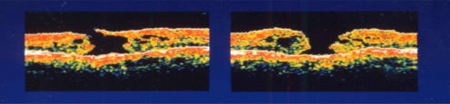
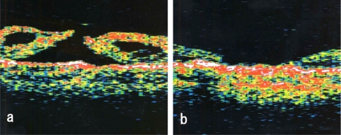
Preoperative optical coherence tomography of both eyes in case 1. Left figure shows right eye (stage III) and right figure is left eye (stage IV).
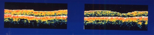
Postoperative optical coherence tomography in case 1. Left figure is the right eye and the right figure is the left eye. Both holes are closed.
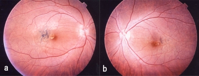
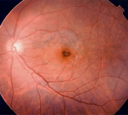
Postoperative fundus photograph of case 1. A yellow round degenerative lesion is seen in the fovea. Irregular degeneration foci associated with pigment deposition can be seen just temporal to the lesion. (a, right eye; b, left eye).
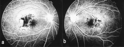
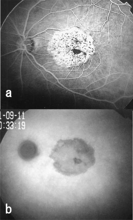
Fluorescein angiogram taken after vitrectomy shows hypofluorescence or blockage of dye in the area with pigment epithelial proliferation in case 1. Hyperfluorescence is noted in the area surrounding the pigment cell proliferation. Many tiny hypofluorescent spots are seen in these areas. (a, right eye; b, left eye).
Optical coherence tomograms of left eye of Case 2. a: Preoperative optical coherence tomogram showing stage IV macular hole. b: Postoperative optical coherence tomogram showing that the macular hole is closed.
Postoperative fundus photograph showing hyperpigmentation around the macular hole and degeneration of pigment the epithelium within the peeled area of internal limiting membrane in case 2.
Fluoresceint angiogram and indocyanine green angiogram of the left eye of Case 2. a: Postoperative fluorescein angiogram showing hypofluorescence and hyperfluorescence areas. b: Postoperative indocyanine green angiogram showing hypofluorescence in the area where ILM was peeled.
Similar articles
-
Idiopathic macular hole surgery in Chinese patients: a randomised study to compare indocyanine green-assisted internal limiting membrane peeling with no internal limiting membrane peeling.Hong Kong Med J. 2005 Aug;11(4):259-66. Hong Kong Med J. 2005. PMID: 16085942 Clinical Trial.
-
Long-term follow-up of indocyanine green-assisted peeling of the retinal internal limiting membrane during vitrectomy surgery for idiopathic macular hole repair.Ophthalmology. 2004 Dec;111(12):2246-53. doi: 10.1016/j.ophtha.2004.05.037. Ophthalmology. 2004. PMID: 15582081
-
Retinal pigment epithelial changes after macular hole surgery with indocyanine green-assisted internal limiting membrane peeling.Am J Ophthalmol. 2002 Jan;133(1):89-94. doi: 10.1016/s0002-9394(01)01293-4. Am J Ophthalmol. 2002. PMID: 11755843
-
Value of internal limiting membrane peeling in surgery for idiopathic macular hole and the correlation between function and retinal morphology.Acta Ophthalmol. 2009 Dec;87 Thesis 2:1-23. doi: 10.1111/j.1755-3768.2009.01777.x. Acta Ophthalmol. 2009. PMID: 19912135 Clinical Trial.
-
Controversies over the role of internal limiting membrane peeling during vitrectomy in macular hole surgery.Surv Ophthalmol. 2017 Jan-Feb;62(1):58-69. doi: 10.1016/j.survophthal.2016.07.003. Epub 2016 Aug 1. Surv Ophthalmol. 2017. PMID: 27491476 Review.
Cited by
-
Retinal pigment epithelium atrophy after epiretinal membrane and internal limiting membrane peeling: case reports.Rom J Ophthalmol. 2022 Jan-Mar;66(1):79-83. doi: 10.22336/rjo.2022.16. Rom J Ophthalmol. 2022. PMID: 35531456 Free PMC article.
References
-
- Smiddy WE, Pimentel S, Williams GA. Macular hole surgery without using adjunctive additives. Ophthalmic Surg Lasers. 1997;28:713–7. - PubMed
-
- Brooks HL Jr. Macular hole surgery with and without internal limiting membrane peeling. Ophthalmology. 2000;107:1939–49. - PubMed
-
- Tognetto D, Grandin R, Sanguinetti G, et al. Internal limiting membrane removal during macular hole surgery – Results of a multicenter retrospective study. Ophthalmology. 2006;113:1401–10. - PubMed
-
- Olsen TW, Sternberg P Jr, Capone A Jr, et al. Macular hole surgery using thrombin-activated fibrinogen and selective removal of the internal limiting membrane. Retina. 1998;18:322–9. - PubMed
-
- Park SS, Marcus DM, Duker JS, et al. Posterior segment complications after vitrectomy for macular hole. Ophthalmology. 1995;102:775–81. - PubMed
LinkOut - more resources
Full Text Sources