. 2009 May 29;4(5):e5735.
doi: 10.1371/journal.pone.0005735.

Selective killing of tumors deficient in methylthioadenosine phosphorylase: a novel strategy

Affiliations

Selective killing of tumors deficient in methylthioadenosine phosphorylase: a novel strategy

Martin Lubin et al. PLoS One. .

Erratum in

  • PLoS One. 2010;5(4). doi: 10.1371/annotation/54fad81d-c975-4b30-bb2b-249650ec3d66

Abstract

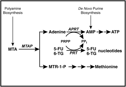
Background: The gene for methylthioadenosine phosphorylase (MTAP) lies on 9p21, close to the gene CDKN2A that encodes the tumor suppressor proteins p16 and p14ARF. MTAP and CDKN2A are homozygously co-deleted, with a frequency of 35 to 70%, in lung and pancreatic cancer, glioblastoma, osteosarcoma, soft-tissue sarcoma, mesothelioma, and T-cell acute lymphoblastic leukemia. In normal cells, but not in tumor cells lacking MTAP, MTAP cleaves the natural substrate, 5'-deoxy-5'-methylthioadenosine (MTA), to adenine and 5-methylthioribose-1-phosphate (MTR-1-P), which are then converted to adenine nucleotides and methionine. This distinct difference between normal MTAP-positive cells and tumor MTAP-negative cells led to several proposals for therapy. We offer a novel strategy in which both MTA and a toxic adenine analog, such as 2,6-diaminopurine (DAP), 6-methylpurine (MeP), or 2-fluoroadenine (F-Ade), are administered. In MTAP-positive cells, abundant adenine, generated from supplied MTA, competitively blocks the conversion of an analog, by adenine phosphoribosyltransferase (APRT), to its active nucleotide form. In MTAP-negative tumor cells, the supplied MTA cannot generate adenine; hence conversion of the analog is not blocked.

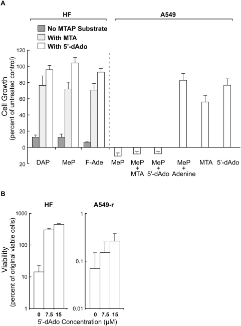
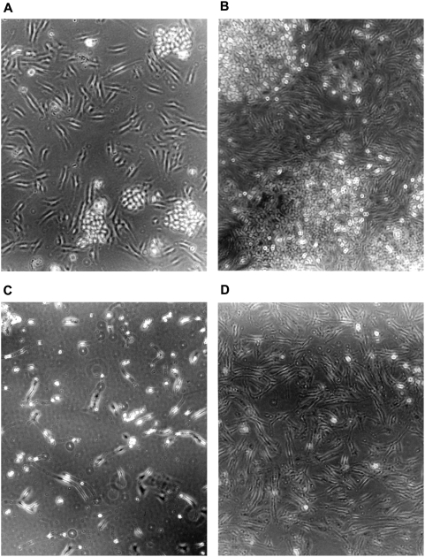
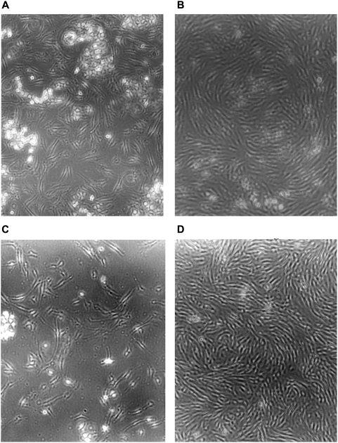
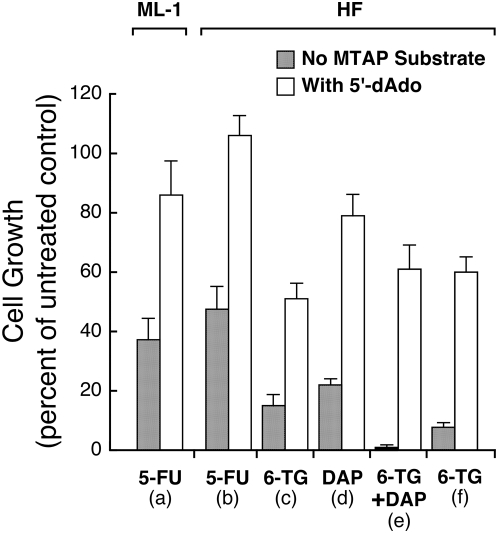
Principal findings: We show that this combination treatment--adenine analog plus MTA--kills MTAP-negative A549 lung tumor cells, while MTAP-positive human fibroblasts (HF) are protected. In co-cultures of the breast tumor cell line, MCF-7, and HF cells, MCF-7 is inhibited or killed, while HF cells proliferate robustly. 5-Fluorouracil (5-FU) and 6-thioguanine (6-TG) may also be used with our strategy. Though neither analog is activated by APRT, in MTAP-positive cells, adenine produced from supplied MTA blocks conversion of 5-FU and 6-TG to their toxic nucleotide forms by competing for 5-phosphoribosyl-1-pyrophosphate (PRPP). The combination of MTA with 5-FU or 6-TG, in the treatment of MTAP-negative tumors, may produce a significantly improved therapeutic index.

Conclusion: We describe a selective strategy to kill tumor cells lacking MTAP.


Conflict of interest statement

Competing Interests: The authors have applied for a patent based on the work presented.

Figures

Figure 1
Figure 1. MTAP metabolic pathway.
In normal cells, MTAP cleaves MTA, a by-product of polyamine biosynthesis, into adenine and MTR-1-P. Adenine is converted to AMP by the ubiquitous enzyme APRT, with PRPP serving as donor of the phosphoribosyl group. MTR-1-P is converted by a series of steps to methionine. AMP is also produced in cells by de novo purine biosynthesis. In addition to APRT, other cellular phosphoribosyltransferases (PRTs), such as hypoxanthine-guanine phsophoribosyl transferase and orotate phosphoribosyltranferase, covert purines and pyrimidines to nucleotides.
Figure 2
Figure 2. MTA and 5′-dAdo protect MTAP-positive HF, but not MTAP-negative A549 cells, from toxic adenine analogs.
(A) HF cells were cultured with the adenine analogs DAP (100 µM), MeP (5 µM), and F-Ade (0.3 µM) with either MTA (15 µM) or 5′-dAdo (15 µM) for three days. Dishes were then trypsinized and cell numbers measured with a Coulter counter. Data from three independent experiments, bars SE. MTAP-negative A549 cells were cultured for three days with MeP (5 µM) and either MTA (15 µM), 5′-dAdo (15 µM), or adenine (15 µM). As controls, cells were cultured with MTA alone (15 µM) or 5′-dAdo alone (15 µM). Dishes were then trypsinized and cell numbers measured with a Coulter counter. Data from three independent experiments, bars SE. (B) For co-culture experiments, the ouabain-resistant cell line A549-r was used. Stocks of HF and A549-r cells were trypsinized and cell numbers counted to determine the amount of each cell line to be seeded for co-culture. To measure the initial number of viable cells, each cell line was separately seeded, in duplicate, into dishes for clonogenic assay. For co-cultures, each dish had 45,000 A549-r and 3,000 HF cells, with DAP at 125 µM, and 5′-dAdo at 0, 7.5, or 15 µM. After three days of incubation, two dishes were trypsinized and serially diluted into fresh medium, with adenine (15 µM) added to suppress further action of DAP, for clonogenic assay of viable A549-r clones. After two additional days of culture, ouabain (0.1 µM) was added to these dishes to inhibit or kill HF cells, while permitting the small number of surviving A540-r cells to grow and form large clones. The remaining two dishes did not receive ouabain and hence viable HF clones could grow and be identified by staining. Counts from duplicate dishes were averaged. Data from two independent experiments, bars SE.
Figure 3
Figure 3. Strategy applied to co-culture of MTAP-negative MCF-7 and MTAP-positive HF cells.
MCF-7 and HF cells were co-cultured with drugs added as follows: (A) Start of co-culture. (B) Three days without drugs. (C) Three days with DAP (200 µM). (D) Three days with DAP (200 µM) and MTA (50 µM). Representative fields are shown.
Figure 4
Figure 4. Strategy with prolonged subculture applied to co-culture of MTAP-negative MCF-7 and MTAP-positive HF cells.
MCF-7 and HF cells were co-cultured with drugs added as follows: (A) Start of co-culture. (B) Four days with DAP (100 µM) and MTA (25 µM). (C) Co-culture in panel B split 1:5 into drug-free medium and fixed two days later. (D) Same as in panel C except culture fixed after five days. Representative fields are shown.
Figure 5
Figure 5. Protection by 5′-dAdo from toxicity of 5-FU and 6-TG in MTAP-positive ML-1 and HF cells.
(a) ML-1 cells were cultured in RPMI 1640 with 10% FBS, for three days, with 5-FU (3 µM), with or without 5′-dAdo (60 µM). Cell numbers were then measured with a Coulter counter. Data from three independent experiments, bars SE. (b) HF cells were cultured in DMEM with 10% FBS for four days, with 5-FU (3 µM), with or without 5′-dAdo (60 µM). Dishes were then trypsinized and cell numbers measured. Data from three independent experiments, bars SE. (c, d, e) HF cells were cultured in DMEM with 10% FBS for three days with either 6-TG (20 µM), DAP (50 µM), or both, with or without 5′-dAdo (15 µM). Dishes were then trypsinized and cell numbers measured with a Coulter counter. Data from three independent experiments, bars SE. (f) HF cells were cultured in DMEM with 10% FBS for three days with 6-TG (40 µM), with or without 5′-dAdo (15 µM). Data from three independent experiments, bars SE.

References

    1. Pegg AE, Williams-Ashman HG. Phosphate-stimulated breakdown of 5′-methylthioadenosine by rat ventral prostate. Biochem J. 1969;115:241–247. - PMC - PubMed
    1. Williams-Ashman HG, Seidenfeld J, Galletti P. Trends in the biochemical pharmacology of 5′-deoxy-5′-methylthioadenosine. Biochem Pharmacol. 1982;31:277–288. - PubMed
    1. Toohey JI. Methylthioadenosine nucleoside phosphorylase deficiency in methylthio-dependent cancer cells. Biochem Biophys Res Commun. 1978;83:27–35. - PubMed
    1. Nobori T, Szinai I, Amox D, Parker B, Olopade OI, et al. Methylthioadenosine phosphorylase deficiency in human non-small cell lung cancers. Cancer Res. 1993;53:1098–1101. - PubMed
    1. Caldas C, Hahn SA, da Costa LT, Redston MS, Schutte M, et al. Frequent somatic mutations and homozygous deletions of the p16 (MTS1) gene in pancreatic adenocarcinoma. Nat Genet. 1994;8:27–32. - PubMed